

changes in the trimeric complex [Cu 3 (3,5-(CF 3 ) 2 pyrazolate) 3 ] [116]. This system
differs from the examples discussed above because the photoexcitation occurs
intermolecularly between two neighbouring copper trimer molecules, as opposed
to the intramolecular processes in the di-nuclear species. Photoactivation at
λ ¼ 355 nm and 17 K promotes the production of excited state species with
microsecond lifetimes, involving a rearrangement of the trimer molecules into
pairs such that one inter-planar Cu. . .Cu distance is reduced by 0.65 Å while the
next Cu. . .Cu contact lengthens by ca. 0.30 Å. In a separate study, conducted in
2009, significant structural distortions in the complex [Cu(dmp)(dppe)](PF 6 ),
(dmp ¼ 2,9-dimethyl-1,10-phenanthroline) are described [117]. The complex crystallises with two, crystallographically independent molecules in the asymmetric unit,
whose structural response on photoactivation is interestingly different. These differences have been attributed to different constraining effects from the surrounding
crystalline environment for each of the independent molecules. In general, upon
irradiation the Cu cation is observed to “flatten out”, and a concomitant increase in
the average Cu–P bond length is observed by comparison of the diffraction data from
the ground and excited states (Fig. 8). The changes are expected to be the result of
charge-transfer between the dmp and dppe ligands and were determined from an
excited state population of ca. 7–10% in the single crystal.
These studies highlighted the importance of the crystalline environment on the
solid-state photoactivation process and confirmed the significance of the “reaction
cavity” hypothesis discussed earlier [97, 118]. In order to exploit this aspect and to
use it to increase the level of conversion to the excited state species, photoactive
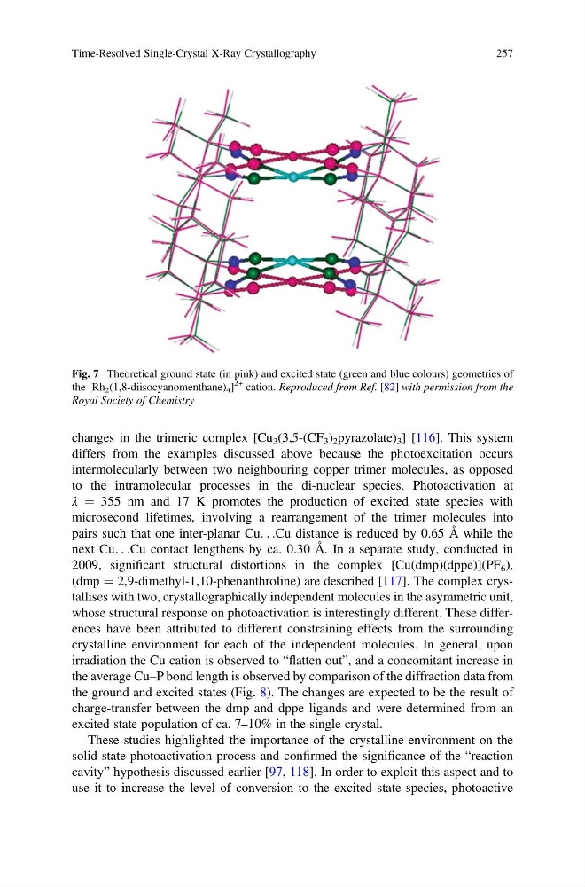
Fig. 7 Theoretical ground state (in pink) and excited state (green and blue colours) geometries of
the [Rh 2 (1,8-diisocyanomenthane) 4 ]
2+ cation. Reproduced from Ref. [82] with permission from the
Royal Society of Chemistry
Time-Resolved Single-Crystal X-Ray Crystallography
257
differs from the examples discussed above because the photoexcitation occurs
intermolecularly between two neighbouring copper trimer molecules, as opposed
to the intramolecular processes in the di-nuclear species. Photoactivation at
λ ¼ 355 nm and 17 K promotes the production of excited state species with
microsecond lifetimes, involving a rearrangement of the trimer molecules into
pairs such that one inter-planar Cu. . .Cu distance is reduced by 0.65 Å while the
next Cu. . .Cu contact lengthens by ca. 0.30 Å. In a separate study, conducted in
2009, significant structural distortions in the complex [Cu(dmp)(dppe)](PF 6 ),
(dmp ¼ 2,9-dimethyl-1,10-phenanthroline) are described [117]. The complex crystallises with two, crystallographically independent molecules in the asymmetric unit,
whose structural response on photoactivation is interestingly different. These differences have been attributed to different constraining effects from the surrounding
crystalline environment for each of the independent molecules. In general, upon
irradiation the Cu cation is observed to “flatten out”, and a concomitant increase in
the average Cu–P bond length is observed by comparison of the diffraction data from
the ground and excited states (Fig. 8). The changes are expected to be the result of
charge-transfer between the dmp and dppe ligands and were determined from an
excited state population of ca. 7–10% in the single crystal.
These studies highlighted the importance of the crystalline environment on the
solid-state photoactivation process and confirmed the significance of the “reaction
cavity” hypothesis discussed earlier [97, 118]. In order to exploit this aspect and to
use it to increase the level of conversion to the excited state species, photoactive
Fig. 7 Theoretical ground state (in pink) and excited state (green and blue colours) geometries of
the [Rh 2 (1,8-diisocyanomenthane) 4 ]
2+ cation. Reproduced from Ref. [82] with permission from the
Royal Society of Chemistry
Time-Resolved Single-Crystal X-Ray Crystallography
257