

58
P. Lecoq
3.1.3.4 Medical Imaging
Scintillators are widely used in medical imaging for X-ray radiology (digital
radiography and CT scanners) and for emission tomography (PET and SPECT) with
a market exceeding several hundred tons per year (see Sect. 20.1).
The choice of the scintillator for medical imaging devices is determined by the
stopping power for the energy range of X and γ-rays to be considered, or more
precisely the conversion efficiency. Materials with high Z and high density are
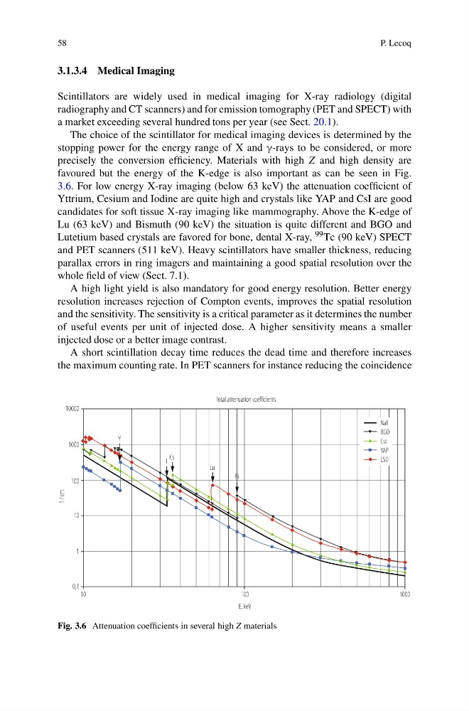
favoured but the energy of the K-edge is also important as can be seen in Fig.
3.6. For low energy X-ray imaging (below 63 keV) the attenuation coefficient of
Yttrium, Cesium and Iodine are quite high and crystals like YAP and CsI are good
candidates for soft tissue X-ray imaging like mammography. Above the K-edge of
Lu (63 keV) and Bismuth (90 keV) the situation is quite different and BGO and
Lutetium based crystals are favored for bone, dental X-ray, 99 Tc (90 keV) SPECT
and PET scanners (511 keV). Heavy scintillators have smaller thickness, reducing
parallax errors in ring imagers and maintaining a good spatial resolution over the
whole field of view (Sect. 7.1).
A high light yield is also mandatory for good energy resolution. Better energy
resolution increases rejection of Compton events, improves the spatial resolution
and the sensitivity. The sensitivity is a critical parameter as it determines the number
of useful events per unit of injected dose. A higher sensitivity means a smaller
injected dose or a better image contrast.
A short scintillation decay time reduces the dead time and therefore increases
the maximum counting rate. In PET scanners for instance reducing the coincidence
Fig. 3.6 Attenuation coefficients in several high Z materials
P. Lecoq
3.1.3.4 Medical Imaging
Scintillators are widely used in medical imaging for X-ray radiology (digital
radiography and CT scanners) and for emission tomography (PET and SPECT) with
a market exceeding several hundred tons per year (see Sect. 20.1).
The choice of the scintillator for medical imaging devices is determined by the
stopping power for the energy range of X and γ-rays to be considered, or more
precisely the conversion efficiency. Materials with high Z and high density are
favoured but the energy of the K-edge is also important as can be seen in Fig.
3.6. For low energy X-ray imaging (below 63 keV) the attenuation coefficient of
Yttrium, Cesium and Iodine are quite high and crystals like YAP and CsI are good
candidates for soft tissue X-ray imaging like mammography. Above the K-edge of
Lu (63 keV) and Bismuth (90 keV) the situation is quite different and BGO and
Lutetium based crystals are favored for bone, dental X-ray, 99 Tc (90 keV) SPECT
and PET scanners (511 keV). Heavy scintillators have smaller thickness, reducing
parallax errors in ring imagers and maintaining a good spatial resolution over the
whole field of view (Sect. 7.1).
A high light yield is also mandatory for good energy resolution. Better energy
resolution increases rejection of Compton events, improves the spatial resolution
and the sensitivity. The sensitivity is a critical parameter as it determines the number
of useful events per unit of injected dose. A higher sensitivity means a smaller
injected dose or a better image contrast.
A short scintillation decay time reduces the dead time and therefore increases
the maximum counting rate. In PET scanners for instance reducing the coincidence
Fig. 3.6 Attenuation coefficients in several high Z materials