

9 Nuclear Emulsions
431
1
a
0.5
tanθ
y
tanθ x
0
0
0.5
1
0
1
2
3
4
5
6
7
8
9
–0.5
–0.5
–1 –1
1
b
0.5
tanθ
y
tanθ x
0
0
0.5
1
0
1
2
3
4
5
6
7
8
9
–0.5
–0.5
–1 –1
New void
B
A
B
A
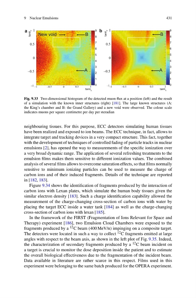
Fig. 9.33 Two-dimensional histogram of the detected muon flux at a position (left) and the result
of a simulation with the known inner structures (right) [181]. The large known structures (A:
the King’s chamber and B: the Grand Gallery) and a new void were observed. The colour scale
indicates muons per square centimetre per day per steradian
neighbouring tissues. For this purpose, ECC detectors simulating human tissues
have been realized and exposed to ion beams. The ECC technique, in fact, allows to
integrate target and tracking devices in a very compact structure. This fact, together
with the development of techniques of controlled fading of particle tracks in nuclear
emulsions [2], has opened the way to measurements of the specific ionization over
a very broad dynamic range. The application of several refreshing treatments to the
emulsion films makes them sensitive to different ionization values. The combined
analysis of several films allows to overcome saturation effects, so that films normally
sensitive to minimum ionizing particles can be used to measure the charge of
carbon ions and of their induced fragments. Details of the technique are reported
in [182, 183].
Figure 9.34 shows the identification of fragments produced by the interaction of
carbon ions with Lexan plates, which simulate the human body tissues given the
similar electron density [183]. Such a charge identification capability allowed the
measurement of the charge-changing cross-section of carbon ions with water by
placing the target ECC inside a water tank [184] as well as the charge-changing
cross-section of carbon ions with lexan [185].
In the framework of the FIRST (Fragmentation of Ions Relevant for Space and
Therapy) experiment [186], two Emulsion Cloud Chambers were exposed to the
fragments produced by a 12 C beam (400 MeV/n) impinging on a composite target.
The detectors were located in such a way to collect 12 C fragments emitted at large
angles with respect to the beam axis, as shown in the left plot of Fig. 9.35. Indeed,
the characterization of secondary fragments produced by a 12 C beam incident on
a target is crucial to monitor the dose deposition inside the patient and to estimate
the overall biological effectiveness due to the fragmentation of the incident beam.
Data available in literature are rather scarce in this respect. Films used in this
experiment were belonging to the same batch produced for the OPERA experiment.
431
1
a
0.5
tanθ
y
tanθ x
0
0
0.5
1
0
1
2
3
4
5
6
7
8
9
–0.5
–0.5
–1 –1
1
b
0.5
tanθ
y
tanθ x
0
0
0.5
1
0
1
2
3
4
5
6
7
8
9
–0.5
–0.5
–1 –1
New void
B
A
B
A
Fig. 9.33 Two-dimensional histogram of the detected muon flux at a position (left) and the result
of a simulation with the known inner structures (right) [181]. The large known structures (A:
the King’s chamber and B: the Grand Gallery) and a new void were observed. The colour scale
indicates muons per square centimetre per day per steradian
neighbouring tissues. For this purpose, ECC detectors simulating human tissues
have been realized and exposed to ion beams. The ECC technique, in fact, allows to
integrate target and tracking devices in a very compact structure. This fact, together
with the development of techniques of controlled fading of particle tracks in nuclear
emulsions [2], has opened the way to measurements of the specific ionization over
a very broad dynamic range. The application of several refreshing treatments to the
emulsion films makes them sensitive to different ionization values. The combined
analysis of several films allows to overcome saturation effects, so that films normally
sensitive to minimum ionizing particles can be used to measure the charge of
carbon ions and of their induced fragments. Details of the technique are reported
in [182, 183].
Figure 9.34 shows the identification of fragments produced by the interaction of
carbon ions with Lexan plates, which simulate the human body tissues given the
similar electron density [183]. Such a charge identification capability allowed the
measurement of the charge-changing cross-section of carbon ions with water by
placing the target ECC inside a water tank [184] as well as the charge-changing
cross-section of carbon ions with lexan [185].
In the framework of the FIRST (Fragmentation of Ions Relevant for Space and
Therapy) experiment [186], two Emulsion Cloud Chambers were exposed to the
fragments produced by a 12 C beam (400 MeV/n) impinging on a composite target.
The detectors were located in such a way to collect 12 C fragments emitted at large
angles with respect to the beam axis, as shown in the left plot of Fig. 9.35. Indeed,
the characterization of secondary fragments produced by a 12 C beam incident on
a target is crucial to monitor the dose deposition inside the patient and to estimate
the overall biological effectiveness due to the fragmentation of the incident beam.
Data available in literature are rather scarce in this respect. Films used in this
experiment were belonging to the same batch produced for the OPERA experiment.