

430
A. Ariga et al.
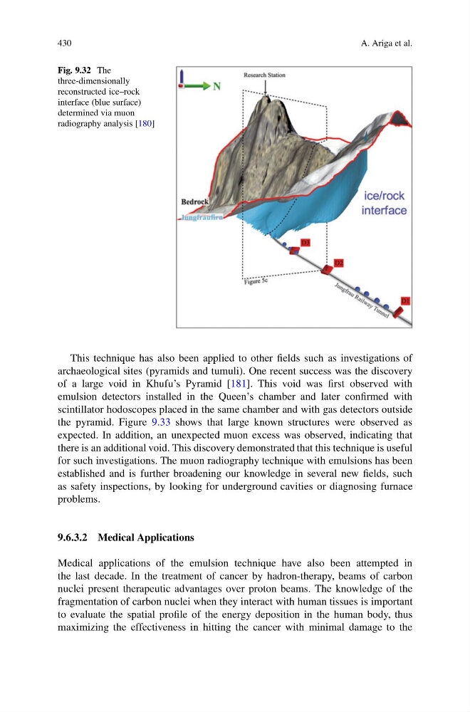
Fig. 9.32 The
three-dimensionally
reconstructed ice–rock
interface (blue surface)
determined via muon
radiography analysis [180]
This technique has also been applied to other fields such as investigations of
archaeological sites (pyramids and tumuli). One recent success was the discovery
of a large void in Khufu’s Pyramid [181]. This void was first observed with
emulsion detectors installed in the Queen’s chamber and later confirmed with
scintillator hodoscopes placed in the same chamber and with gas detectors outside
the pyramid. Figure 9.33 shows that large known structures were observed as
expected. In addition, an unexpected muon excess was observed, indicating that
there is an additional void. This discovery demonstrated that this technique is useful
for such investigations. The muon radiography technique with emulsions has been
established and is further broadening our knowledge in several new fields, such
as safety inspections, by looking for underground cavities or diagnosing furnace
problems.
9.6.3.2 Medical Applications
Medical applications of the emulsion technique have also been attempted in
the last decade. In the treatment of cancer by hadron-therapy, beams of carbon
nuclei present therapeutic advantages over proton beams. The knowledge of the
fragmentation of carbon nuclei when they interact with human tissues is important
to evaluate the spatial profile of the energy deposition in the human body, thus
maximizing the effectiveness in hitting the cancer with minimal damage to the
A. Ariga et al.
Fig. 9.32 The
three-dimensionally
reconstructed ice–rock
interface (blue surface)
determined via muon
radiography analysis [180]
This technique has also been applied to other fields such as investigations of
archaeological sites (pyramids and tumuli). One recent success was the discovery
of a large void in Khufu’s Pyramid [181]. This void was first observed with
emulsion detectors installed in the Queen’s chamber and later confirmed with
scintillator hodoscopes placed in the same chamber and with gas detectors outside
the pyramid. Figure 9.33 shows that large known structures were observed as
expected. In addition, an unexpected muon excess was observed, indicating that
there is an additional void. This discovery demonstrated that this technique is useful
for such investigations. The muon radiography technique with emulsions has been
established and is further broadening our knowledge in several new fields, such
as safety inspections, by looking for underground cavities or diagnosing furnace
problems.
9.6.3.2 Medical Applications
Medical applications of the emulsion technique have also been attempted in
the last decade. In the treatment of cancer by hadron-therapy, beams of carbon
nuclei present therapeutic advantages over proton beams. The knowledge of the
fragmentation of carbon nuclei when they interact with human tissues is important
to evaluate the spatial profile of the energy deposition in the human body, thus
maximizing the effectiveness in hitting the cancer with minimal damage to the