

6.4 Crystallinity and Phase Identity of Powdered Samples
165
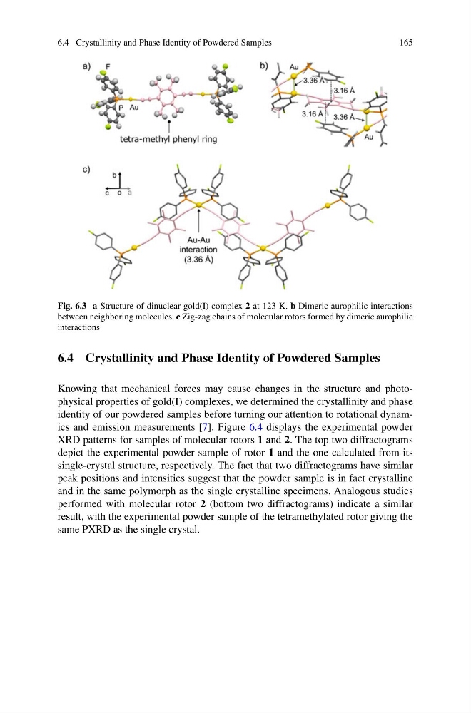
Fig. 6.3 a Structure of dinuclear gold(I) complex 2 at 123 K. b Dimeric aurophilic interactions
between neighboring molecules. c Zig-zag chains of molecular rotors formed by dimeric aurophilic
interactions
6.4 Crystallinity and Phase Identity of Powdered Samples
Knowing that mechanical forces may cause changes in the structure and photophysical properties of gold(I) complexes, we determined the crystallinity and phase
identity of our powdered samples before turning our attention to rotational dynamics and emission measurements [7]. Figure 6.4 displays the experimental powder
XRD patterns for samples of molecular rotors 1 and 2. The top two diffractograms
depict the experimental powder sample of rotor 1 and the one calculated from its
single-crystal structure, respectively. The fact that two diffractograms have similar
peak positions and intensities suggest that the powder sample is in fact crystalline
and in the same polymorph as the single crystalline specimens. Analogous studies
performed with molecular rotor 2 (bottom two diffractograms) indicate a similar
result, with the experimental powder sample of the tetramethylated rotor giving the
same PXRD as the single crystal.
165
Fig. 6.3 a Structure of dinuclear gold(I) complex 2 at 123 K. b Dimeric aurophilic interactions
between neighboring molecules. c Zig-zag chains of molecular rotors formed by dimeric aurophilic
interactions
6.4 Crystallinity and Phase Identity of Powdered Samples
Knowing that mechanical forces may cause changes in the structure and photophysical properties of gold(I) complexes, we determined the crystallinity and phase
identity of our powdered samples before turning our attention to rotational dynamics and emission measurements [7]. Figure 6.4 displays the experimental powder
XRD patterns for samples of molecular rotors 1 and 2. The top two diffractograms
depict the experimental powder sample of rotor 1 and the one calculated from its
single-crystal structure, respectively. The fact that two diffractograms have similar
peak positions and intensities suggest that the powder sample is in fact crystalline
and in the same polymorph as the single crystalline specimens. Analogous studies
performed with molecular rotor 2 (bottom two diffractograms) indicate a similar
result, with the experimental powder sample of the tetramethylated rotor giving the
same PXRD as the single crystal.