

(e.g., InAs/CdSe or InAs/CdSe/ZnSe) leading to a dramatically higher quantum
yield (10 times to Ag 2 Se QDs) and size-tunable emission in NIR-II region. In this
study, they demonstrated that these QDs functionalized via three distinct surface
coatings (e.g., triglyceride-rich lipoproteins, phospholipid micelles, PEG2000 PE)
can allow for functional imaging to measure metabolic rates of lipoproteins in
several organs and heartbeat/breathing rates as well as to quantify the blood flow
of mouse brain vasculature [22].
Despite all of these photo-physical advantages of QDs, a substantial challenge for
conventional Cd-containing QDs is their inherent cytotoxicity [23]. For example,
CdSe QDs were found to induce cell death due to the liberated free Cd
2+ from the
CdSe lattice. Therefore, many studies have been performed to develop Cd-free QDs
(e.g., InP, InAs, Ag) with comparable or even better performance than existing
Cd-containing QDs [24]. Surface passivation via an additional inorganic shell
(e.g., ZnS capping of CdSe QDs) is another way of reducing the oxidation-mediated
cytotoxicity of Cd-QDs; here, PL improvement also appears owing to the effective
passivation of surface non-radiative recombination of excitons [25]. Nevertheless,
besides the aforementioned composition controls, toxicity issues can be
circumvented by size control of QDs to make them renally clearable. In a pioneering
study using QDs as a model inorganic nanoparticle, Frangioni and coworkers
proposed the design considerations to reduce metal-induced toxicity [26]. In this
study, it was revealed that nanoparticles of hydrodynamic diameter <5.5 nm could
be efficiently excreted in urine via systemic intravenously injections.
3 Anti-Stokes Shift Luminescent Nanoparticles
Anti-Stokes shift luminescence is a special optical process of converting (low
energy) long wavelength of light to (higher-energy) short-wavelength radiation,
which can permit the deeper tissue penetration with minimal photo-bleaching
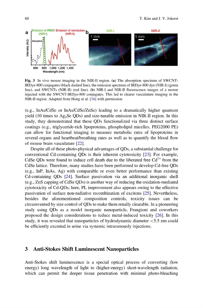
Fig. 3 In vivo mouse imaging in the NIR-II region. (a) The absorption spectrum of SWCNTIRDye-800 conjugates (black dashed line), the emission spectrum of IRDye-800 dye (NIR-I) (green
line), and SWCNTs (NIR-II) (red line). (b) NIR-I and NIR-II fluorescence images of a mouse
injected with the SWCNT-IRDye-800 conjugates. This led to clearer vasculature imaging in the
NIR-II region. Adapted from Hong et al. [16] with permission
60
T. Kim and J. V. Jokerst
yield (10 times to Ag 2 Se QDs) and size-tunable emission in NIR-II region. In this
study, they demonstrated that these QDs functionalized via three distinct surface
coatings (e.g., triglyceride-rich lipoproteins, phospholipid micelles, PEG2000 PE)
can allow for functional imaging to measure metabolic rates of lipoproteins in
several organs and heartbeat/breathing rates as well as to quantify the blood flow
of mouse brain vasculature [22].
Despite all of these photo-physical advantages of QDs, a substantial challenge for
conventional Cd-containing QDs is their inherent cytotoxicity [23]. For example,
CdSe QDs were found to induce cell death due to the liberated free Cd
2+ from the
CdSe lattice. Therefore, many studies have been performed to develop Cd-free QDs
(e.g., InP, InAs, Ag) with comparable or even better performance than existing
Cd-containing QDs [24]. Surface passivation via an additional inorganic shell
(e.g., ZnS capping of CdSe QDs) is another way of reducing the oxidation-mediated
cytotoxicity of Cd-QDs; here, PL improvement also appears owing to the effective
passivation of surface non-radiative recombination of excitons [25]. Nevertheless,
besides the aforementioned composition controls, toxicity issues can be
circumvented by size control of QDs to make them renally clearable. In a pioneering
study using QDs as a model inorganic nanoparticle, Frangioni and coworkers
proposed the design considerations to reduce metal-induced toxicity [26]. In this
study, it was revealed that nanoparticles of hydrodynamic diameter <5.5 nm could
be efficiently excreted in urine via systemic intravenously injections.
3 Anti-Stokes Shift Luminescent Nanoparticles
Anti-Stokes shift luminescence is a special optical process of converting (low
energy) long wavelength of light to (higher-energy) short-wavelength radiation,
which can permit the deeper tissue penetration with minimal photo-bleaching
Fig. 3 In vivo mouse imaging in the NIR-II region. (a) The absorption spectrum of SWCNTIRDye-800 conjugates (black dashed line), the emission spectrum of IRDye-800 dye (NIR-I) (green
line), and SWCNTs (NIR-II) (red line). (b) NIR-I and NIR-II fluorescence images of a mouse
injected with the SWCNT-IRDye-800 conjugates. This led to clearer vasculature imaging in the
NIR-II region. Adapted from Hong et al. [16] with permission
60
T. Kim and J. V. Jokerst