

4.5 Fluorescence Prodrug Conjugates
Fluorescence prodrug conjugates are dual functional systems that offer both
therapeutic promise and potential for concurrent diagnosis, which are able to target
cancer cells selectively, provide cytotoxic chemotherapeutics, and allow facile
monitoring of the location and efficacy of drugs [109, 110]. Fluorescence prodrug
conjugates are of particular interest since they are stable in blood plasma, which can
be activated efficiently by various cellular constituents, such as thiols, reactive
oxygen species (ROS), and enzymes that are overexpressed in tumors [111, 112].
Fluorescence prodrug conjugates can realize both targeted therapeutic release and
targeted FMI. Such prodrug conjugates usually contain fluorophores, cleavable
linkers, and targeting ligands or chemotherapeutic agents. Fluorophores are usually
naphthalimide, coumarin, BODIPY, rhodol, cyanine, etc. Cleavage linkers include
hydrolysis of esters, amides, and hydrazine linkers, disulfide exchange-based
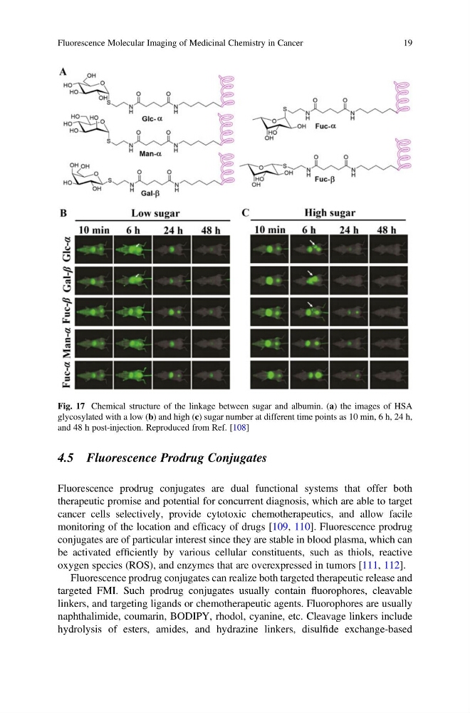
Fig. 17 Chemical structure of the linkage between sugar and albumin. (a) the images of HSA
glycosylated with a low (b) and high (c) sugar number at different time points as 10 min, 6 h, 24 h,
and 48 h post-injection. Reproduced from Ref. [108]
Fluorescence Molecular Imaging of Medicinal Chemistry in Cancer
19
Fluorescence prodrug conjugates are dual functional systems that offer both
therapeutic promise and potential for concurrent diagnosis, which are able to target
cancer cells selectively, provide cytotoxic chemotherapeutics, and allow facile
monitoring of the location and efficacy of drugs [109, 110]. Fluorescence prodrug
conjugates are of particular interest since they are stable in blood plasma, which can
be activated efficiently by various cellular constituents, such as thiols, reactive
oxygen species (ROS), and enzymes that are overexpressed in tumors [111, 112].
Fluorescence prodrug conjugates can realize both targeted therapeutic release and
targeted FMI. Such prodrug conjugates usually contain fluorophores, cleavable
linkers, and targeting ligands or chemotherapeutic agents. Fluorophores are usually
naphthalimide, coumarin, BODIPY, rhodol, cyanine, etc. Cleavage linkers include
hydrolysis of esters, amides, and hydrazine linkers, disulfide exchange-based
Fig. 17 Chemical structure of the linkage between sugar and albumin. (a) the images of HSA
glycosylated with a low (b) and high (c) sugar number at different time points as 10 min, 6 h, 24 h,
and 48 h post-injection. Reproduced from Ref. [108]
Fluorescence Molecular Imaging of Medicinal Chemistry in Cancer
19