

growth with treatment for a period of time. However, this method can only tell the
changes in tumors when they show anatomical changes; in addition, it is difficult to
evaluate the effect of in situ tumor therapy by the traditional method. FMI can
completely overcome the shortcomings of the traditional method and can not only
monitor the changes of the tumor biomarker but also evaluate the therapeutic effect
in an early and dynamic manner. For example, histone deacetylases (HDACs) are
overexpressed in TNBC. The FMI of the LBH589-Cy5.5 probe has been successfully applied not only for measuring the expression and functions of HDACs in
tumors but also in evaluating the therapeutic response of HDAC inhibitor SAHA
treatment, as evidenced by the significantly reduced HDAC signals in SAHA-treated
breast tumors (Fig. 15) [106].
4.3 Tracking the Drug Biodistribution and Metabolism
When the tumor cells were treated with drugs, it was difficult to observe the drug
interaction with the corresponding targets by traditional pharmacochemical methods.
The majority of drugs tested clinically exhibit off-target effects, which is easy to
produce side effects. FMI is able to directly visualize the binding of the drug to the
target, which can effectively improve the success rate of drug development. For
example, 2-((3-(3-fluoro-4-hydroxyphenyl)-7-hydroxynaphthalen-1-yl) methylene)
malononitrile (FPNM) can potently inhibit the growth of MDA-MB-231 tumors, and
the relative binding affinity (RBA) value shows FPNM is an estrogen receptor β
(ERβ) ligand. In order to confirm the interaction between FPNM and ERβ, FMI of
FPNM is performed in MCF-7 cells. As shown in Fig. 16, ERβ is a nuclear hormone
receptor, and the fluorescence derived from the complex between FPNM and ERβ
was mainly detected in the cell nucleus. The data suggested that FPNM showed
specifically selective affinity toward ERβ in the living MCF-7 cells. These results
indicated that FPNM possesses the ability to selectively bind to the ERβ in living
cells [107].
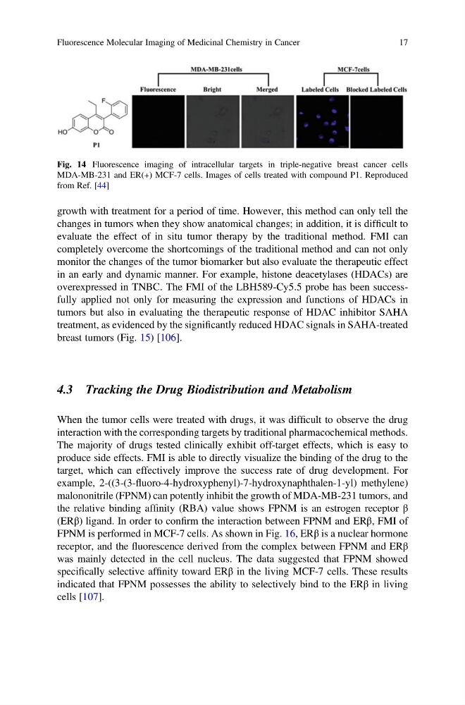
Fig. 14 Fluorescence imaging of intracellular targets in triple-negative breast cancer cells
MDA-MB-231 and ER(+) MCF-7 cells. Images of cells treated with compound P1. Reproduced
from Ref. [44]
Fluorescence Molecular Imaging of Medicinal Chemistry in Cancer
17
changes in tumors when they show anatomical changes; in addition, it is difficult to
evaluate the effect of in situ tumor therapy by the traditional method. FMI can
completely overcome the shortcomings of the traditional method and can not only
monitor the changes of the tumor biomarker but also evaluate the therapeutic effect
in an early and dynamic manner. For example, histone deacetylases (HDACs) are
overexpressed in TNBC. The FMI of the LBH589-Cy5.5 probe has been successfully applied not only for measuring the expression and functions of HDACs in
tumors but also in evaluating the therapeutic response of HDAC inhibitor SAHA
treatment, as evidenced by the significantly reduced HDAC signals in SAHA-treated
breast tumors (Fig. 15) [106].
4.3 Tracking the Drug Biodistribution and Metabolism
When the tumor cells were treated with drugs, it was difficult to observe the drug
interaction with the corresponding targets by traditional pharmacochemical methods.
The majority of drugs tested clinically exhibit off-target effects, which is easy to
produce side effects. FMI is able to directly visualize the binding of the drug to the
target, which can effectively improve the success rate of drug development. For
example, 2-((3-(3-fluoro-4-hydroxyphenyl)-7-hydroxynaphthalen-1-yl) methylene)
malononitrile (FPNM) can potently inhibit the growth of MDA-MB-231 tumors, and
the relative binding affinity (RBA) value shows FPNM is an estrogen receptor β
(ERβ) ligand. In order to confirm the interaction between FPNM and ERβ, FMI of
FPNM is performed in MCF-7 cells. As shown in Fig. 16, ERβ is a nuclear hormone
receptor, and the fluorescence derived from the complex between FPNM and ERβ
was mainly detected in the cell nucleus. The data suggested that FPNM showed
specifically selective affinity toward ERβ in the living MCF-7 cells. These results
indicated that FPNM possesses the ability to selectively bind to the ERβ in living
cells [107].
Fig. 14 Fluorescence imaging of intracellular targets in triple-negative breast cancer cells
MDA-MB-231 and ER(+) MCF-7 cells. Images of cells treated with compound P1. Reproduced
from Ref. [44]
Fluorescence Molecular Imaging of Medicinal Chemistry in Cancer
17