

4.1 Identification of Therapeutic Targets
Specific therapeutic target is the key for therapy, but traditional drug chemistry
methods find targets at slow speed. FMI can improve the process of target identification and identify suitable treatment regimens, hence improving patient treatment
outcomes. For example, breast cancer is the most common cancer among women
with different subtypes. Nearly 75% of patients demonstrate abnormally high
expression of ERα. Therefore, ERα is regarded as an important pharmaceutical
target for the treatment of breast cancer, and many ERα ligands have been developed
into hormone agents. However, hormone therapy is ineffective for ERα(À) and
triple-negative breast cancers (TNBCs). The ERα fluorescent probe P1 can be used
to identify a suitable therapeutic regimen for breast cancer. As shown in Fig. 14, the
fluorescence signals can be observed in the cell nucleus of the ERα-positive MCF-7
breast cancer cell, but not in MDA-MB-231 TNBC cells. Therefore, FMI is able to
identify the target expression and determine treatment [45].
4.2 Candidate Drug Screening
To evaluate the therapeutic effects of antitumor drugs in vivo, a traditional medical
imaging method is used to measure the tumor volume at the late stage of tumor
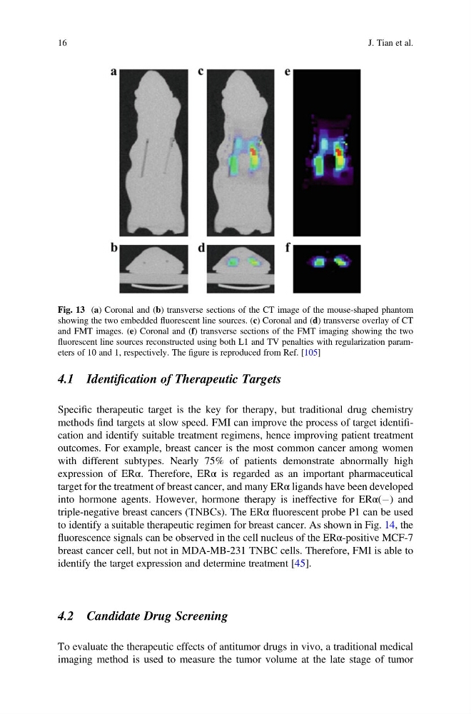
Fig. 13 (a) Coronal and (b) transverse sections of the CT image of the mouse-shaped phantom
showing the two embedded fluorescent line sources. (c) Coronal and (d) transverse overlay of CT
and FMT images. (e) Coronal and (f) transverse sections of the FMT imaging showing the two
fluorescent line sources reconstructed using both L1 and TV penalties with regularization parameters of 10 and 1, respectively. The figure is reproduced from Ref. [105]
16
J. Tian et al.
Specific therapeutic target is the key for therapy, but traditional drug chemistry
methods find targets at slow speed. FMI can improve the process of target identification and identify suitable treatment regimens, hence improving patient treatment
outcomes. For example, breast cancer is the most common cancer among women
with different subtypes. Nearly 75% of patients demonstrate abnormally high
expression of ERα. Therefore, ERα is regarded as an important pharmaceutical
target for the treatment of breast cancer, and many ERα ligands have been developed
into hormone agents. However, hormone therapy is ineffective for ERα(À) and
triple-negative breast cancers (TNBCs). The ERα fluorescent probe P1 can be used
to identify a suitable therapeutic regimen for breast cancer. As shown in Fig. 14, the
fluorescence signals can be observed in the cell nucleus of the ERα-positive MCF-7
breast cancer cell, but not in MDA-MB-231 TNBC cells. Therefore, FMI is able to
identify the target expression and determine treatment [45].
4.2 Candidate Drug Screening
To evaluate the therapeutic effects of antitumor drugs in vivo, a traditional medical
imaging method is used to measure the tumor volume at the late stage of tumor
Fig. 13 (a) Coronal and (b) transverse sections of the CT image of the mouse-shaped phantom
showing the two embedded fluorescent line sources. (c) Coronal and (d) transverse overlay of CT
and FMT images. (e) Coronal and (f) transverse sections of the FMT imaging showing the two
fluorescent line sources reconstructed using both L1 and TV penalties with regularization parameters of 10 and 1, respectively. The figure is reproduced from Ref. [105]
16
J. Tian et al.