

could undergo 1,6-elimination of p-quinone methide to generate a
tetraphenylethylene-pyridinium compound with poor solubility, resulting in the
appearance of aggregation effect and a light-up fluorescence of the TPE moiety
based on the AIE effect. The TPE-Gal displayed distinguishable advantages such as
high specificity toward β-gal, no self-quenching at high concentration, large Stokes
shift and so on. Moreover, the researchers also tested the applicability of TPE-Gal
for sensing β-gal not only in aqueous solution but also in living cells. The results
showed that TPE-Gal could exhibit excellent fluorescence properties in these environments (Fig. 5).
3 Nanomaterial for β-Galactosidase Activity Detection
The nanomaterial has attracted considerable attentions from the early 1980s due to
its special physical and chemical properties and promising application. Entering the
twenty-first century, the researches of nanomaterials have been extended in various
fields, especially in chemical sensing and biomedical imaging such as the detection
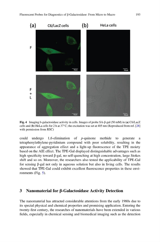
Fig. 4 Imaging b-galactosidase activity in cells. Images of probe SA-β-gal (50 mM) in (a) C6/LacZ
cells and (b) HeLa cells for 2 h at 37
C; the excitation was set at 405 nm (Reproduced from ref. [28]
with permission from RSC)
Fluorescent Probes for Diagnostics of β-Galactosidase: From Micro to Macro
193
tetraphenylethylene-pyridinium compound with poor solubility, resulting in the
appearance of aggregation effect and a light-up fluorescence of the TPE moiety
based on the AIE effect. The TPE-Gal displayed distinguishable advantages such as
high specificity toward β-gal, no self-quenching at high concentration, large Stokes
shift and so on. Moreover, the researchers also tested the applicability of TPE-Gal
for sensing β-gal not only in aqueous solution but also in living cells. The results
showed that TPE-Gal could exhibit excellent fluorescence properties in these environments (Fig. 5).
3 Nanomaterial for β-Galactosidase Activity Detection
The nanomaterial has attracted considerable attentions from the early 1980s due to
its special physical and chemical properties and promising application. Entering the
twenty-first century, the researches of nanomaterials have been extended in various
fields, especially in chemical sensing and biomedical imaging such as the detection
Fig. 4 Imaging b-galactosidase activity in cells. Images of probe SA-β-gal (50 mM) in (a) C6/LacZ
cells and (b) HeLa cells for 2 h at 37
C; the excitation was set at 405 nm (Reproduced from ref. [28]
with permission from RSC)
Fluorescent Probes for Diagnostics of β-Galactosidase: From Micro to Macro
193