

segments which can stretch out to a great extent, giving the thread its elasticity.
Mechanical forces (such as tension) not only are a stress to cells but play a central
role in many developmental, physiological, and pathological processes, especially
regarding the transduction of signals. One of the exciting results in this field was
produced by Grashoff et al., who have designed a tension sensor module (TSMod) to
examine the mechanical forces across vinculin during cell migration [158]. In this
sensor, a 40-amino-acid-long elastic domain was inserted between two fluorophores
(mTFP1 and Venus (A206K)) as a potential fluorescence resonance energy transfer
(FRET) pair. The elastic domain derived from the spider silk protein flagelliform
consists of repetitive amino acid motifs that form entropic nanosprings suitable
for measuring piconewton forces. Since FRET is highly sensitive to the distance
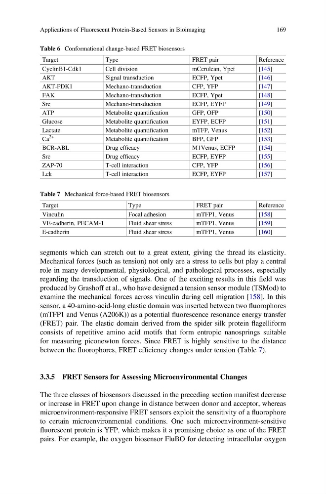
between the fluorophores, FRET efficiency changes under tension (Table 7).
3.3.5 FRET Sensors for Assessing Microenvironmental Changes
The three classes of biosensors discussed in the preceding section manifest decrease
or increase in FRET upon change in distance between donor and acceptor, whereas
microenvironment-responsive FRET sensors exploit the sensitivity of a fluorophore
to certain microenvironmental conditions. One such microenvironment-sensitive
fluorescent protein is YFP, which makes it a promising choice as one of the FRET
pairs. For example, the oxygen biosensor FluBO for detecting intracellular oxygen
Table 6 Conformational change-based FRET biosensors
Target
Type
FRET pair
Reference
CyclinB1-Cdk1
Cell division
mCerulean, Ypet
[145]
AKT
Signal transduction
ECFP, Ypet
[146]
AKT-PDK1
Mechano-transduction
CFP, YFP
[147]
FAK
Mechano-transduction
ECFP, Ypet
[148]
Src
Mechano-transduction
ECFP, EYFP
[149]
ATP
Metabolite quantification
GFP, OFP
[150]
Glucose
Metabolite quantification
EYFP, ECFP
[151]
Lactate
Metabolite quantification
mTFP, Venus
[152]
Ca
2+
Metabolite quantification
BFP, GFP
[153]
BCR-ABL
Drug efficacy
M1Venus, ECFP
[154]
Src
Drug efficacy
ECFP, EYFP
[155]
ZAP-70
T-cell interaction
CFP, YFP
[156]
Lck
T-cell interaction
ECFP, EYFP
[157]
Table 7 Mechanical force-based FRET biosensors
Target
Type
FRET pair
Reference
Vinculin
Focal adhesion
mTFP1, Venus
[158]
VE-cadherin, PECAM-1
Fluid shear stress
mTFP1, Venus
[159]
E-cadherin
Fluid shear stress
mTFP1, Venus
[160]
Applications of Fluorescent Protein-Based Sensors in Bioimaging
169
Mechanical forces (such as tension) not only are a stress to cells but play a central
role in many developmental, physiological, and pathological processes, especially
regarding the transduction of signals. One of the exciting results in this field was
produced by Grashoff et al., who have designed a tension sensor module (TSMod) to
examine the mechanical forces across vinculin during cell migration [158]. In this
sensor, a 40-amino-acid-long elastic domain was inserted between two fluorophores
(mTFP1 and Venus (A206K)) as a potential fluorescence resonance energy transfer
(FRET) pair. The elastic domain derived from the spider silk protein flagelliform
consists of repetitive amino acid motifs that form entropic nanosprings suitable
for measuring piconewton forces. Since FRET is highly sensitive to the distance
between the fluorophores, FRET efficiency changes under tension (Table 7).
3.3.5 FRET Sensors for Assessing Microenvironmental Changes
The three classes of biosensors discussed in the preceding section manifest decrease
or increase in FRET upon change in distance between donor and acceptor, whereas
microenvironment-responsive FRET sensors exploit the sensitivity of a fluorophore
to certain microenvironmental conditions. One such microenvironment-sensitive
fluorescent protein is YFP, which makes it a promising choice as one of the FRET
pairs. For example, the oxygen biosensor FluBO for detecting intracellular oxygen
Table 6 Conformational change-based FRET biosensors
Target
Type
FRET pair
Reference
CyclinB1-Cdk1
Cell division
mCerulean, Ypet
[145]
AKT
Signal transduction
ECFP, Ypet
[146]
AKT-PDK1
Mechano-transduction
CFP, YFP
[147]
FAK
Mechano-transduction
ECFP, Ypet
[148]
Src
Mechano-transduction
ECFP, EYFP
[149]
ATP
Metabolite quantification
GFP, OFP
[150]
Glucose
Metabolite quantification
EYFP, ECFP
[151]
Lactate
Metabolite quantification
mTFP, Venus
[152]
Ca
2+
Metabolite quantification
BFP, GFP
[153]
BCR-ABL
Drug efficacy
M1Venus, ECFP
[154]
Src
Drug efficacy
ECFP, EYFP
[155]
ZAP-70
T-cell interaction
CFP, YFP
[156]
Lck
T-cell interaction
ECFP, EYFP
[157]
Table 7 Mechanical force-based FRET biosensors
Target
Type
FRET pair
Reference
Vinculin
Focal adhesion
mTFP1, Venus
[158]
VE-cadherin, PECAM-1
Fluid shear stress
mTFP1, Venus
[159]
E-cadherin
Fluid shear stress
mTFP1, Venus
[160]
Applications of Fluorescent Protein-Based Sensors in Bioimaging
169