

determine the activation of specific proteases, often following the stimulation of
a pathway. For example, in the case of the caspase-3 biosensor, in the presence
of active enzyme, the single-peptide sensor DEVD (caspase cleavage sequence) is
cleaved, increasing the distance between CFP and eYFP, resulting in increased CFP
fluorescence and decreased FRET [136]. Another major limitation is that measuring
loss of signal is a readout for measuring FRET rather than measuring an increase in
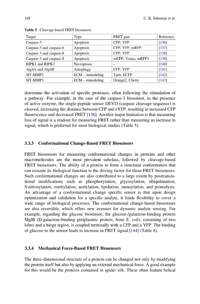
signal, which is preferred for most biological studies (Table 5).
3.3.3 Conformational Change-Based FRET Biosensors
FRET biosensors for measuring conformational changes in proteins and other
macromolecules are the most prevalent subclass, followed by cleavage-based
FRET biosensors. The ability of a protein to form a structural conformation that
can execute its biological function is the driving factor for these FRET biosensors.
Such conformational changes are also contributed to a large extent by posttranslational modifications such as phosphorylation, glycosylation, ubiquitination,
S-nitrosylation, methylation, acetylation, lipidation, sumoylation, and proteolysis.
An advantage of a conformational change specific sensor is that upon design
optimization and validation for a specific analyte, it lends flexibility to cover a
wide range of biological processes. The conformational change-based biosensors
are also reversible, which offers new avenues for dynamic analyte sensing. For
example, regarding the glucose biosensor, the glucose-/galactose-binding protein
MglB (D-galactose-binding periplasmic protein, from E. coli), consisting of two
lobes and a hinge region, is coupled terminally with a CFP and a YFP. The binding
of glucose to the sensor leads to increase in FRET signal [144] (Table 6).
3.3.4 Mechanical Force-Based FRET Biosensors
The three-dimensional structure of a protein can be changed not only by modifying
the protein itself but also by applying an external mechanical force. A good example
for this would be the proteins contained in spider silk. These often feature helical
Table 5 Cleavage-based FRET biosensors
Target
Type
FRET pair
Reference
Caspase-3
Apoptosis
CFP, YFP
[136]
Caspase-3 and caspase-6
Apoptosis
CFP, YFP, mRFP
[137]
Caspase-3 and caspase-8
Apoptosis
CFP, YFP
[138]
Caspase-3 and caspase-8
Apoptosis
seCFP, Venus, mRFP1
[139]
RIPK1 and RIPK3
Necroptosis
–
[140]
Atg4A and Atg4B
Autophagy
CFP, YFP
[141]
MT-MMP1
ECM – remodeling
Ypet, ECFP
[142]
MT-MMP1
ECM – remodeling
Orange2, Cherry
[143]
168
U. K. Sukumar et al.
a pathway. For example, in the case of the caspase-3 biosensor, in the presence
of active enzyme, the single-peptide sensor DEVD (caspase cleavage sequence) is
cleaved, increasing the distance between CFP and eYFP, resulting in increased CFP
fluorescence and decreased FRET [136]. Another major limitation is that measuring
loss of signal is a readout for measuring FRET rather than measuring an increase in
signal, which is preferred for most biological studies (Table 5).
3.3.3 Conformational Change-Based FRET Biosensors
FRET biosensors for measuring conformational changes in proteins and other
macromolecules are the most prevalent subclass, followed by cleavage-based
FRET biosensors. The ability of a protein to form a structural conformation that
can execute its biological function is the driving factor for these FRET biosensors.
Such conformational changes are also contributed to a large extent by posttranslational modifications such as phosphorylation, glycosylation, ubiquitination,
S-nitrosylation, methylation, acetylation, lipidation, sumoylation, and proteolysis.
An advantage of a conformational change specific sensor is that upon design
optimization and validation for a specific analyte, it lends flexibility to cover a
wide range of biological processes. The conformational change-based biosensors
are also reversible, which offers new avenues for dynamic analyte sensing. For
example, regarding the glucose biosensor, the glucose-/galactose-binding protein
MglB (D-galactose-binding periplasmic protein, from E. coli), consisting of two
lobes and a hinge region, is coupled terminally with a CFP and a YFP. The binding
of glucose to the sensor leads to increase in FRET signal [144] (Table 6).
3.3.4 Mechanical Force-Based FRET Biosensors
The three-dimensional structure of a protein can be changed not only by modifying
the protein itself but also by applying an external mechanical force. A good example
for this would be the proteins contained in spider silk. These often feature helical
Table 5 Cleavage-based FRET biosensors
Target
Type
FRET pair
Reference
Caspase-3
Apoptosis
CFP, YFP
[136]
Caspase-3 and caspase-6
Apoptosis
CFP, YFP, mRFP
[137]
Caspase-3 and caspase-8
Apoptosis
CFP, YFP
[138]
Caspase-3 and caspase-8
Apoptosis
seCFP, Venus, mRFP1
[139]
RIPK1 and RIPK3
Necroptosis
–
[140]
Atg4A and Atg4B
Autophagy
CFP, YFP
[141]
MT-MMP1
ECM – remodeling
Ypet, ECFP
[142]
MT-MMP1
ECM – remodeling
Orange2, Cherry
[143]
168
U. K. Sukumar et al.