

3.3.1 Types of FRET Biosensors
The intramolecular FRET probes/sensors involve two fluorescently tagged proteins
that bring them into close proximity in the event of an interaction between the two
analytes to generate FRET signal. In the following section, we also classify FRET
biosensors by their way of transforming a biological change into a change in FRET
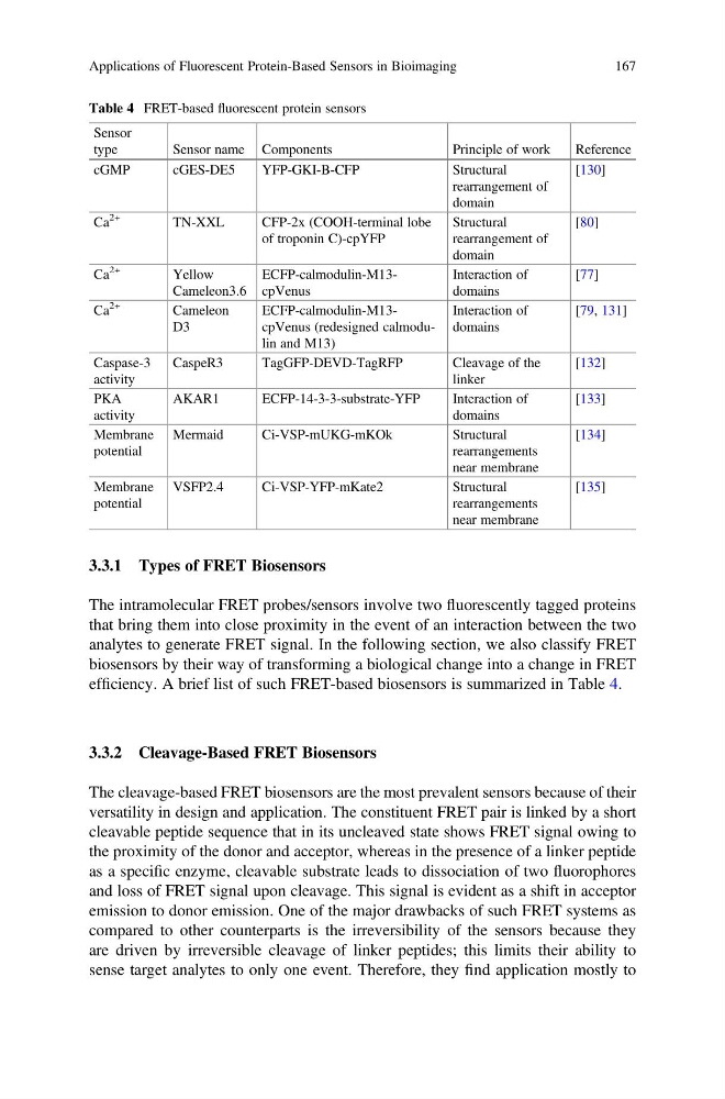
efficiency. A brief list of such FRET-based biosensors is summarized in Table 4.
3.3.2 Cleavage-Based FRET Biosensors
The cleavage-based FRET biosensors are the most prevalent sensors because of their
versatility in design and application. The constituent FRET pair is linked by a short
cleavable peptide sequence that in its uncleaved state shows FRET signal owing to
the proximity of the donor and acceptor, whereas in the presence of a linker peptide
as a specific enzyme, cleavable substrate leads to dissociation of two fluorophores
and loss of FRET signal upon cleavage. This signal is evident as a shift in acceptor
emission to donor emission. One of the major drawbacks of such FRET systems as
compared to other counterparts is the irreversibility of the sensors because they
are driven by irreversible cleavage of linker peptides; this limits their ability to
sense target analytes to only one event. Therefore, they find application mostly to
Table 4 FRET-based fluorescent protein sensors
Sensor
type
Sensor name Components
Principle of work
Reference
cGMP
cGES-DE5
YFP-GKI-B-CFP
Structural
rearrangement of
domain
[130]
Ca
2+
TN-XXL
CFP-2x (COOH-terminal lobe
of troponin C)-cpYFP
Structural
rearrangement of
domain
[80]
Ca
2+
Yellow
Cameleon3.6
ECFP-calmodulin-M13cpVenus
Interaction of
domains
[77]
Ca
2+
Cameleon
D3
ECFP-calmodulin-M13cpVenus (redesigned calmodulin and M13)
Interaction of
domains
[79, 131]
Caspase-3
activity
CaspeR3
TagGFP-DEVD-TagRFP
Cleavage of the
linker
[132]
PKA
activity
AKAR1
ECFP-14-3-3-substrate-YFP
Interaction of
domains
[133]
Membrane
potential
Mermaid
Ci-VSP-mUKG-mKOk
Structural
rearrangements
near membrane
[134]
Membrane
potential
VSFP2.4
Ci-VSP-YFP-mKate2
Structural
rearrangements
near membrane
[135]
Applications of Fluorescent Protein-Based Sensors in Bioimaging
167
The intramolecular FRET probes/sensors involve two fluorescently tagged proteins
that bring them into close proximity in the event of an interaction between the two
analytes to generate FRET signal. In the following section, we also classify FRET
biosensors by their way of transforming a biological change into a change in FRET
efficiency. A brief list of such FRET-based biosensors is summarized in Table 4.
3.3.2 Cleavage-Based FRET Biosensors
The cleavage-based FRET biosensors are the most prevalent sensors because of their
versatility in design and application. The constituent FRET pair is linked by a short
cleavable peptide sequence that in its uncleaved state shows FRET signal owing to
the proximity of the donor and acceptor, whereas in the presence of a linker peptide
as a specific enzyme, cleavable substrate leads to dissociation of two fluorophores
and loss of FRET signal upon cleavage. This signal is evident as a shift in acceptor
emission to donor emission. One of the major drawbacks of such FRET systems as
compared to other counterparts is the irreversibility of the sensors because they
are driven by irreversible cleavage of linker peptides; this limits their ability to
sense target analytes to only one event. Therefore, they find application mostly to
Table 4 FRET-based fluorescent protein sensors
Sensor
type
Sensor name Components
Principle of work
Reference
cGMP
cGES-DE5
YFP-GKI-B-CFP
Structural
rearrangement of
domain
[130]
Ca
2+
TN-XXL
CFP-2x (COOH-terminal lobe
of troponin C)-cpYFP
Structural
rearrangement of
domain
[80]
Ca
2+
Yellow
Cameleon3.6
ECFP-calmodulin-M13cpVenus
Interaction of
domains
[77]
Ca
2+
Cameleon
D3
ECFP-calmodulin-M13cpVenus (redesigned calmodulin and M13)
Interaction of
domains
[79, 131]
Caspase-3
activity
CaspeR3
TagGFP-DEVD-TagRFP
Cleavage of the
linker
[132]
PKA
activity
AKAR1
ECFP-14-3-3-substrate-YFP
Interaction of
domains
[133]
Membrane
potential
Mermaid
Ci-VSP-mUKG-mKOk
Structural
rearrangements
near membrane
[134]
Membrane
potential
VSFP2.4
Ci-VSP-YFP-mKate2
Structural
rearrangements
near membrane
[135]
Applications of Fluorescent Protein-Based Sensors in Bioimaging
167