

2.1.2 NIR-I Imaging Fluorophores
Fluorophores with emission in the NIR region possess less absorption and scatter
from tissues much more efficiently than fluorophores based on visible light, which is
favorable for in vivo imaging with a high signal-to-background ratio (SBR). This
imaging modality also inherited quick feedback, high-resolution, and noninvasive
properties of optical imaging and can be utilized to visualize the real-time dynamics
in living organisms [20–23]. Over the past decade, fluorescence imaging in the first
NIR window (NIR-I, 700–900 nm) has been widely studied in fundamental research
and preclinical/clinical applications, which is partially because of the immediate
availability of a wide range of fluorophores, such as NIR-760, IRDye800CW,
indocyanine green (ICG), methylene blue, and their derivatives (Fig. 4) [24–26].
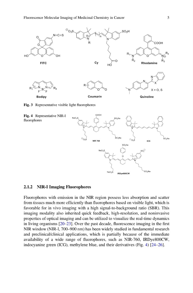
Fig. 3 Representative visible light fluorophores
Fig. 4 Representative NIR-I
fluorophores
Fluorescence Molecular Imaging of Medicinal Chemistry in Cancer
5
Fluorophores with emission in the NIR region possess less absorption and scatter
from tissues much more efficiently than fluorophores based on visible light, which is
favorable for in vivo imaging with a high signal-to-background ratio (SBR). This
imaging modality also inherited quick feedback, high-resolution, and noninvasive
properties of optical imaging and can be utilized to visualize the real-time dynamics
in living organisms [20–23]. Over the past decade, fluorescence imaging in the first
NIR window (NIR-I, 700–900 nm) has been widely studied in fundamental research
and preclinical/clinical applications, which is partially because of the immediate
availability of a wide range of fluorophores, such as NIR-760, IRDye800CW,
indocyanine green (ICG), methylene blue, and their derivatives (Fig. 4) [24–26].
Fig. 3 Representative visible light fluorophores
Fig. 4 Representative NIR-I
fluorophores
Fluorescence Molecular Imaging of Medicinal Chemistry in Cancer
5