

potential use to obtain both anatomic and molecular information through CT and
NIR-II imaging, as shown in Fig. 14 [60].
By now, it is still a great challenge to identify early metastases in cancer
diagnostics and therapy. Here, Moghe et al. show that intravenously injected
albumin-encapsulated NIR-II RENPs can detect targeted metastatic lesions
in vivo, allowing for the longitudinal tracking of multi-organ metastases. In a murine
model of human breast cancer, the NIR-II nanoprobes enabled whole-body detection
of adrenal gland microlesions and bone lesions that were not available via contrastenhanced MRI as early up to 5 weeks post-inoculation, respectively (see Fig. 15)
[61]. Noninvasive monitoring of gastrointestinal drug release in vivo is another
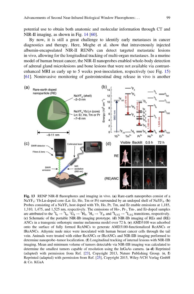
Fig. 13 RENP NIR-II fluorophores and imaging in vivo. (a) Rare-earth nanoprobes consist of a
NaYF 4 : Yb:Ln-doped core (Ln: Er, Ho, Tm or Pr) surrounded by an undoped shell of NaYF 4 . (b)
Probes consisting of a NaYF 4 host doped with Yb, Ho, Pr, Tm, and Er enable emissions at 1,185,
1,310, 1,475, and 1,525 nm, respectively. The emissions of Ho-, Pr-, Tm-, and Er-doped samples
are attributed to the
5
I 6 !
5
I 8 ,
1
G 4 !
3
H 5 ,
3
H 4 !
3
F 4 , and
4
I 13/2 !
4
I 15/2 transitions, respectively.
(c) Schematic of the portable NIR-IIb imaging prototype. (d) NIR-IIb imaging of REs and (RE)
ANCs in a transgenic orthotopic murine melanoma model over 72 h. (e) AMD3100 was adsorbed
onto the surface of fully formed ReANCs to generate AMD3100-functionalized ReANCs or
fReANCs. Athymic nude mice were inoculated with human breast cancer cells through the tail
vein. Animals were treated with either ReANCs or fReANCs and NIR-IIB imaging performed to
determine nanoprobe–tumor localization. (f) Longitudinal tracking of internal lesions with NIR-IIB
imaging. Mean and minimum volume of tumors detectable via NIR-IIB imaging was calculated to
determine the smallest tumors capable of resolution using the InGaAs camera. (a–d) Reprinted
(adapted) with permission from Ref. [23], Copyright 2013, Nature Publishing Group. (e, f)
Reprinted (adapted) with permission from Ref. [25], Copyright 2015, Wiley-VCH Verlag GmbH
& Co. KGaA
Advancements of Second Near-Infrared Biological Window Fluorophores:. . .
99
NIR-II imaging, as shown in Fig. 14 [60].
By now, it is still a great challenge to identify early metastases in cancer
diagnostics and therapy. Here, Moghe et al. show that intravenously injected
albumin-encapsulated NIR-II RENPs can detect targeted metastatic lesions
in vivo, allowing for the longitudinal tracking of multi-organ metastases. In a murine
model of human breast cancer, the NIR-II nanoprobes enabled whole-body detection
of adrenal gland microlesions and bone lesions that were not available via contrastenhanced MRI as early up to 5 weeks post-inoculation, respectively (see Fig. 15)
[61]. Noninvasive monitoring of gastrointestinal drug release in vivo is another
Fig. 13 RENP NIR-II fluorophores and imaging in vivo. (a) Rare-earth nanoprobes consist of a
NaYF 4 : Yb:Ln-doped core (Ln: Er, Ho, Tm or Pr) surrounded by an undoped shell of NaYF 4 . (b)
Probes consisting of a NaYF 4 host doped with Yb, Ho, Pr, Tm, and Er enable emissions at 1,185,
1,310, 1,475, and 1,525 nm, respectively. The emissions of Ho-, Pr-, Tm-, and Er-doped samples
are attributed to the
5
I 6 !
5
I 8 ,
1
G 4 !
3
H 5 ,
3
H 4 !
3
F 4 , and
4
I 13/2 !
4
I 15/2 transitions, respectively.
(c) Schematic of the portable NIR-IIb imaging prototype. (d) NIR-IIb imaging of REs and (RE)
ANCs in a transgenic orthotopic murine melanoma model over 72 h. (e) AMD3100 was adsorbed
onto the surface of fully formed ReANCs to generate AMD3100-functionalized ReANCs or
fReANCs. Athymic nude mice were inoculated with human breast cancer cells through the tail
vein. Animals were treated with either ReANCs or fReANCs and NIR-IIB imaging performed to
determine nanoprobe–tumor localization. (f) Longitudinal tracking of internal lesions with NIR-IIB
imaging. Mean and minimum volume of tumors detectable via NIR-IIB imaging was calculated to
determine the smallest tumors capable of resolution using the InGaAs camera. (a–d) Reprinted
(adapted) with permission from Ref. [23], Copyright 2013, Nature Publishing Group. (e, f)
Reprinted (adapted) with permission from Ref. [25], Copyright 2015, Wiley-VCH Verlag GmbH
& Co. KGaA
Advancements of Second Near-Infrared Biological Window Fluorophores:. . .
99