

challenge because of the limited spatial resolution and long scanning time of existing
bioimaging modalities. Zhang et al. report a novel microcarrier that can retain drugs
and withstand the harsh conditions of gastrointestinal tract. Significantly, they can
track the microcarrier fate and semiquantitatively monitor the content of drug
released in vivo in real time by measuring the fluorescence signals in the second
near-infrared window of RENPs with an absorption competition-induced emission
bioimaging system. The microcarriers show a prolonged residence time of up to 72 h
in the gastrointestinal tract, releasing up to 62% of their content [62].
Current NIR-II fluorophore-related in vivo biodetections are only focused on
direct disease lesion or organ bioimaging; it’s still a big challenge to realize the
NIR-II real-time dynamic biosensing. Very recently, a new type of Er
3+ -sensitized
upconversion nanoparticles with both excitation (1,530 nm) and emission
(1,180 nm) located in NIR-II window for in vivo biosensing is developed [63]. Significantly, the microneedle patch sensor for in vivo inflammation dynamic detection
is developed based on the ratiometric fluorescence by combining the effective
NIR-II upconversion emission and H 2 O 2 sensing organic probes under the Fenton
catalysis of Fe
2+ . Due to the large anti-Stokes shifting, low autofluorescence, and
tissue scattering of the NIR-II upconversion luminescence, the inflammation can be
dynamically evaluated in vivo in real time at high resolution (200 Â 200 μm).
RENPs are also attracted to fluorophores utilized for multimodal imaging. For
example, Cheng et al. have reported a strategy to enhance the PAI intensity of
upconverted nanoparticles (UCNPs) by functionalized with photoswitchable
azobenzene-containing poly (acrylic acid) (UCNPs@PAA-Azo) to yield a PAI
signal that is up to six times higher than that of UCNPs without a decay in NIR-II
emission [64]. Using this nanoprobe, NIR-II imaging provides real-time means for
the precise resection of the lymph node (LN) during surgery, where PA imaging
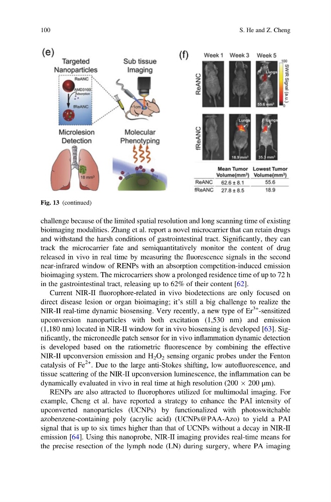
Fig. 13 (continued)
100
S. He and Z. Cheng
bioimaging modalities. Zhang et al. report a novel microcarrier that can retain drugs
and withstand the harsh conditions of gastrointestinal tract. Significantly, they can
track the microcarrier fate and semiquantitatively monitor the content of drug
released in vivo in real time by measuring the fluorescence signals in the second
near-infrared window of RENPs with an absorption competition-induced emission
bioimaging system. The microcarriers show a prolonged residence time of up to 72 h
in the gastrointestinal tract, releasing up to 62% of their content [62].
Current NIR-II fluorophore-related in vivo biodetections are only focused on
direct disease lesion or organ bioimaging; it’s still a big challenge to realize the
NIR-II real-time dynamic biosensing. Very recently, a new type of Er
3+ -sensitized
upconversion nanoparticles with both excitation (1,530 nm) and emission
(1,180 nm) located in NIR-II window for in vivo biosensing is developed [63]. Significantly, the microneedle patch sensor for in vivo inflammation dynamic detection
is developed based on the ratiometric fluorescence by combining the effective
NIR-II upconversion emission and H 2 O 2 sensing organic probes under the Fenton
catalysis of Fe
2+ . Due to the large anti-Stokes shifting, low autofluorescence, and
tissue scattering of the NIR-II upconversion luminescence, the inflammation can be
dynamically evaluated in vivo in real time at high resolution (200 Â 200 μm).
RENPs are also attracted to fluorophores utilized for multimodal imaging. For
example, Cheng et al. have reported a strategy to enhance the PAI intensity of
upconverted nanoparticles (UCNPs) by functionalized with photoswitchable
azobenzene-containing poly (acrylic acid) (UCNPs@PAA-Azo) to yield a PAI
signal that is up to six times higher than that of UCNPs without a decay in NIR-II
emission [64]. Using this nanoprobe, NIR-II imaging provides real-time means for
the precise resection of the lymph node (LN) during surgery, where PA imaging
Fig. 13 (continued)
100
S. He and Z. Cheng