

demonstrate real time, multispectral in vivo NIR-II imaging, which offers anatomical
resolution using a library of tunable and biocompatible RENPs (Fig. 13a–d)
[23]. This work introduces a new generation of versatile NIR-II nanoprobes that
facilitate disease monitoring using minimally invasive NIR-II imaging.
Another novel example reported that DSPE-mPEG-modified rare-earth-doped
nanoparticles (RENPs@DSPE-mPEG) show inherent affinity to bone without
linking any targeting ligands and, thus, provide an alternative noninvasive and
nonradiation strategy for skeletal system mapping and bone disease diagnoses
[59]. Besides bone imaging, RENPs@DSPE-mPEG show an imaging application
in blood vessels and lymph nodes. Importantly, RENPs@DSPE-mPEG can be
internalized by circulating white blood cells. This finding may open a window to
increase efficient nanoparticle delivery in the fields such as immunotherapy and
improve the diagnostic and therapeutic efficacy of cancer-targeted nanoparticles in
clinical applications.
Early tumor detection is crucial for cancer successful treatment. Specific targeting
moieties are key factors for surface functionalization of RENPs. For example,
targeting NIR-II fluorophores have been prepared by encapsulating RENPs with
AMD3100, a hydrophobic small-molecule antagonist of the chemokine receptor
CXCR4. These targeting RENPs preferentially localize to receptor-positive tumors
in mice, allowing detection of CXCR4-positive tumors four times smaller than
receptor-negative tumors (Fig. 13e, f) [25]. These targeted NIR-II fluorophores
enable imaging of microlesions in the lungs at a depth of ~1 cm and are able to
simultaneously identify the phenotype of the tumor population. This study is significant to functionalize NIR-II RENPs to detect sub-tissue microlesions at an early
stage. In another interesting research, RENPs have also been excited by X-ray
instead of laser to produce emission light for NIR-II imaging, highlighting their
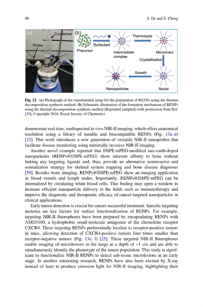
Fig. 12 (a) Photograph of the experimental setup for the preparation of RENPs using the thermal
decomposition synthesis method. (b) Schematic illustration of the formation mechanism of RENPs
using the thermal decomposition synthesis method (Reprinted (adapted) with permission from Ref.
[39], Copyright 2016, Royal Society of Chemistry)
98
S. He and Z. Cheng
resolution using a library of tunable and biocompatible RENPs (Fig. 13a–d)
[23]. This work introduces a new generation of versatile NIR-II nanoprobes that
facilitate disease monitoring using minimally invasive NIR-II imaging.
Another novel example reported that DSPE-mPEG-modified rare-earth-doped
nanoparticles (RENPs@DSPE-mPEG) show inherent affinity to bone without
linking any targeting ligands and, thus, provide an alternative noninvasive and
nonradiation strategy for skeletal system mapping and bone disease diagnoses
[59]. Besides bone imaging, RENPs@DSPE-mPEG show an imaging application
in blood vessels and lymph nodes. Importantly, RENPs@DSPE-mPEG can be
internalized by circulating white blood cells. This finding may open a window to
increase efficient nanoparticle delivery in the fields such as immunotherapy and
improve the diagnostic and therapeutic efficacy of cancer-targeted nanoparticles in
clinical applications.
Early tumor detection is crucial for cancer successful treatment. Specific targeting
moieties are key factors for surface functionalization of RENPs. For example,
targeting NIR-II fluorophores have been prepared by encapsulating RENPs with
AMD3100, a hydrophobic small-molecule antagonist of the chemokine receptor
CXCR4. These targeting RENPs preferentially localize to receptor-positive tumors
in mice, allowing detection of CXCR4-positive tumors four times smaller than
receptor-negative tumors (Fig. 13e, f) [25]. These targeted NIR-II fluorophores
enable imaging of microlesions in the lungs at a depth of ~1 cm and are able to
simultaneously identify the phenotype of the tumor population. This study is significant to functionalize NIR-II RENPs to detect sub-tissue microlesions at an early
stage. In another interesting research, RENPs have also been excited by X-ray
instead of laser to produce emission light for NIR-II imaging, highlighting their
Fig. 12 (a) Photograph of the experimental setup for the preparation of RENPs using the thermal
decomposition synthesis method. (b) Schematic illustration of the formation mechanism of RENPs
using the thermal decomposition synthesis method (Reprinted (adapted) with permission from Ref.
[39], Copyright 2016, Royal Society of Chemistry)
98
S. He and Z. Cheng