

40
2.3.7.3.1 Immobilization of BoNT/A Antibody and SV Protein on CM5 SPR
Sensor Chip
Dhaked and coworkers described the stepwise immobilization of BoNT/A antibody
and SV protein on a CM5 chip, respectively, in nine steps. These steps involved the
stabilization of the baseline, the activation of carboxyl groups on the CM5 chip, and
converting them into activated carboxymethylated groups on the sensor chip for
future bounding to the free amino groups of BoNT/A antibody. This was followed
by washing with PBS and measuring the SPR angle that has shifted nearly to the
baseline [84]. When the BoNT/A antibodies are injected on the CM5 chip, an
increase in the SPR angle was observed. Finally, washing with 1000 mM
ethanolamine was performed to prevent non-specific binding and to block the
unreacted NHS-ester groups on CM5 chip. From Figs. 2.22 and 2.23, a net angle
change of 95.44 m° and 48 m° are observed, and this ascribes the attachment of
0.79 ng/ mm2 of antibody and 0.4 ng/mm2 SV protein on CM5 chip, respectively [84].
2.4 Conclusion
Most of the sensing measuring devices used for the detection of biological warfare
agents are based on luminescence immunoassay signal transduction mechanisms,
which are optical. In this chapter, we have discussed the following subjects: impedance spectroscopy, evanescent wave technology and internal reflection fluorescence
(TIRF) excitation, surface acoustic wave sensors, fluorescent biosensors,
Fluorescence Resonance Energy Transfer (FRET), light emission (CANARY),
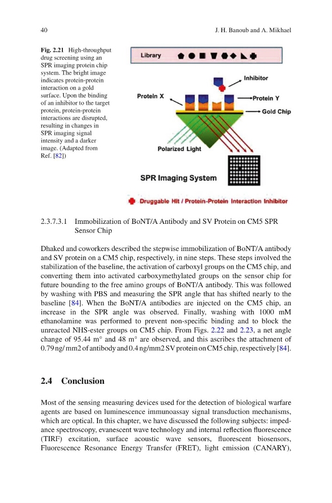
Fig. 2.21 High-throughput
drug screening using an
SPR imaging protein chip
system. The bright image
indicates protein-protein
interaction on a gold
surface. Upon the binding
of an inhibitor to the target
protein, protein-protein
interactions are disrupted,
resulting in changes in
SPR imaging signal
intensity and a darker
image. (Adapted from
Ref. [82])
J. H. Banoub and A. Mikhael
2.3.7.3.1 Immobilization of BoNT/A Antibody and SV Protein on CM5 SPR
Sensor Chip
Dhaked and coworkers described the stepwise immobilization of BoNT/A antibody
and SV protein on a CM5 chip, respectively, in nine steps. These steps involved the
stabilization of the baseline, the activation of carboxyl groups on the CM5 chip, and
converting them into activated carboxymethylated groups on the sensor chip for
future bounding to the free amino groups of BoNT/A antibody. This was followed
by washing with PBS and measuring the SPR angle that has shifted nearly to the
baseline [84]. When the BoNT/A antibodies are injected on the CM5 chip, an
increase in the SPR angle was observed. Finally, washing with 1000 mM
ethanolamine was performed to prevent non-specific binding and to block the
unreacted NHS-ester groups on CM5 chip. From Figs. 2.22 and 2.23, a net angle
change of 95.44 m° and 48 m° are observed, and this ascribes the attachment of
0.79 ng/ mm2 of antibody and 0.4 ng/mm2 SV protein on CM5 chip, respectively [84].
2.4 Conclusion
Most of the sensing measuring devices used for the detection of biological warfare
agents are based on luminescence immunoassay signal transduction mechanisms,
which are optical. In this chapter, we have discussed the following subjects: impedance spectroscopy, evanescent wave technology and internal reflection fluorescence
(TIRF) excitation, surface acoustic wave sensors, fluorescent biosensors,
Fluorescence Resonance Energy Transfer (FRET), light emission (CANARY),
Fig. 2.21 High-throughput
drug screening using an
SPR imaging protein chip
system. The bright image
indicates protein-protein
interaction on a gold
surface. Upon the binding
of an inhibitor to the target
protein, protein-protein
interactions are disrupted,
resulting in changes in
SPR imaging signal
intensity and a darker
image. (Adapted from
Ref. [82])
J. H. Banoub and A. Mikhael