

38
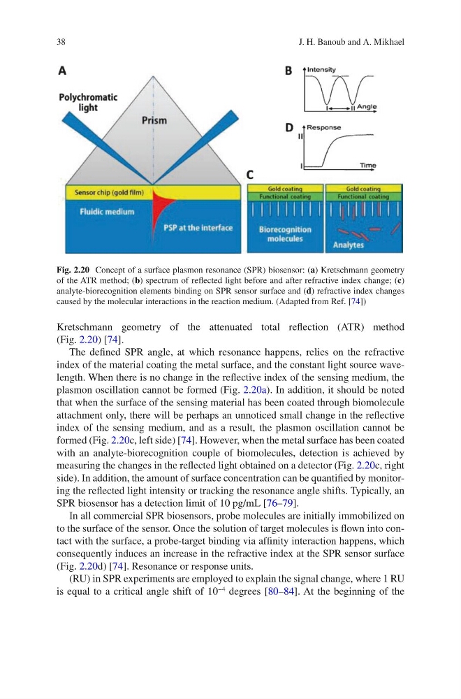
Kretschmann geometry of the attenuated total reflection (ATR) method
(Fig. 2.20) [74].
The defined SPR angle, at which resonance happens, relies on the refractive
index of the material coating the metal surface, and the constant light source wavelength. When there is no change in the reflective index of the sensing medium, the
plasmon oscillation cannot be formed (Fig. 2.20a). In addition, it should be noted
that when the surface of the sensing material has been coated through biomolecule
attachment only, there will be perhaps an unnoticed small change in the reflective
index of the sensing medium, and as a result, the plasmon oscillation cannot be
formed (Fig. 2.20c, left side) [74]. However, when the metal surface has been coated
with an analyte-biorecognition couple of biomolecules, detection is achieved by
measuring the changes in the reflected light obtained on a detector (Fig. 2.20c, right
side). In addition, the amount of surface concentration can be quantified by monitoring the reflected light intensity or tracking the resonance angle shifts. Typically, an
SPR biosensor has a detection limit of 10 pg/mL [76–79].
In all commercial SPR biosensors, probe molecules are initially immobilized on
to the surface of the sensor. Once the solution of target molecules is flown into contact with the surface, a probe-target binding via affinity interaction happens, which
consequently induces an increase in the refractive index at the SPR sensor surface
(Fig. 2.20d) [74]. Resonance or response units.
(RU) in SPR experiments are employed to explain the signal change, where 1 RU
is equal to a critical angle shift of 10
−4
degrees [80–84]. At the beginning of the
Fig. 2.20 Concept of a surface plasmon resonance (SPR) biosensor: (a) Kretschmann geometry
of the ATR method; (b) spectrum of reflected light before and after refractive index change; (c)
analyte-biorecognition elements binding on SPR sensor surface and (d) refractive index changes
caused by the molecular interactions in the reaction medium. (Adapted from Ref. [74])
J. H. Banoub and A. Mikhael
Kretschmann geometry of the attenuated total reflection (ATR) method
(Fig. 2.20) [74].
The defined SPR angle, at which resonance happens, relies on the refractive
index of the material coating the metal surface, and the constant light source wavelength. When there is no change in the reflective index of the sensing medium, the
plasmon oscillation cannot be formed (Fig. 2.20a). In addition, it should be noted
that when the surface of the sensing material has been coated through biomolecule
attachment only, there will be perhaps an unnoticed small change in the reflective
index of the sensing medium, and as a result, the plasmon oscillation cannot be
formed (Fig. 2.20c, left side) [74]. However, when the metal surface has been coated
with an analyte-biorecognition couple of biomolecules, detection is achieved by
measuring the changes in the reflected light obtained on a detector (Fig. 2.20c, right
side). In addition, the amount of surface concentration can be quantified by monitoring the reflected light intensity or tracking the resonance angle shifts. Typically, an
SPR biosensor has a detection limit of 10 pg/mL [76–79].
In all commercial SPR biosensors, probe molecules are initially immobilized on
to the surface of the sensor. Once the solution of target molecules is flown into contact with the surface, a probe-target binding via affinity interaction happens, which
consequently induces an increase in the refractive index at the SPR sensor surface
(Fig. 2.20d) [74]. Resonance or response units.
(RU) in SPR experiments are employed to explain the signal change, where 1 RU
is equal to a critical angle shift of 10
−4
degrees [80–84]. At the beginning of the
Fig. 2.20 Concept of a surface plasmon resonance (SPR) biosensor: (a) Kretschmann geometry
of the ATR method; (b) spectrum of reflected light before and after refractive index change; (c)
analyte-biorecognition elements binding on SPR sensor surface and (d) refractive index changes
caused by the molecular interactions in the reaction medium. (Adapted from Ref. [74])
J. H. Banoub and A. Mikhael