

19
2.3.1.3 Selectivity Measurements
McAlpine also examined the selectivity of the AMP-functionalized biosensors
toward the following various bacterial species (1) Gram-negative pathogenic E. coli
O157∶H7, (2) the nonpathogenic E. coli strain American Type Cell Culture (ATCC)
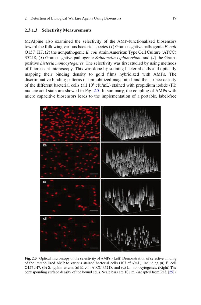
35218, (3) Gram-negative pathogenic Salmonella typhimurium, and (4) the Grampositive Listeria monocytogenes. The selectivity was first studied by using methods
of fluorescent microscopy. This was done by staining bacterial cells and optically
mapping their binding density to gold films hybridized with AMPs. The
discriminative binding patterns of immobilized magainin I and the surface density
of the different bacterial cells (all 10
7
cfu/mL) stained with propidium iodide (PI)
nucleic acid stain are showed in Fig. 2.5. In summary, the coupling of AMPs with
micro capacitive biosensors leads to the implementation of a portable, label-free
Fig. 2.5 Optical microscopy of the selectivity of AMPs. (Left) Demonstration of selective binding
of the immobilized AMP to various stained bacterial cells (107 cfu∕mL), including (a) E. coli
O157∶H7, (b) S. typhimurium, (c) E. coli ATCC 35218, and (d) L. monocytogenes. (Right) The
corresponding surface density of the bound cells. Scale bars are 10 μm. (Adapted from Ref. [25])
2 Detection of Biological Warfare Agents Using Biosensors
2.3.1.3 Selectivity Measurements
McAlpine also examined the selectivity of the AMP-functionalized biosensors
toward the following various bacterial species (1) Gram-negative pathogenic E. coli
O157∶H7, (2) the nonpathogenic E. coli strain American Type Cell Culture (ATCC)
35218, (3) Gram-negative pathogenic Salmonella typhimurium, and (4) the Grampositive Listeria monocytogenes. The selectivity was first studied by using methods
of fluorescent microscopy. This was done by staining bacterial cells and optically
mapping their binding density to gold films hybridized with AMPs. The
discriminative binding patterns of immobilized magainin I and the surface density
of the different bacterial cells (all 10
7
cfu/mL) stained with propidium iodide (PI)
nucleic acid stain are showed in Fig. 2.5. In summary, the coupling of AMPs with
micro capacitive biosensors leads to the implementation of a portable, label-free
Fig. 2.5 Optical microscopy of the selectivity of AMPs. (Left) Demonstration of selective binding
of the immobilized AMP to various stained bacterial cells (107 cfu∕mL), including (a) E. coli
O157∶H7, (b) S. typhimurium, (c) E. coli ATCC 35218, and (d) L. monocytogenes. (Right) The
corresponding surface density of the bound cells. Scale bars are 10 μm. (Adapted from Ref. [25])
2 Detection of Biological Warfare Agents Using Biosensors