

8.8 Early Lung Development
487
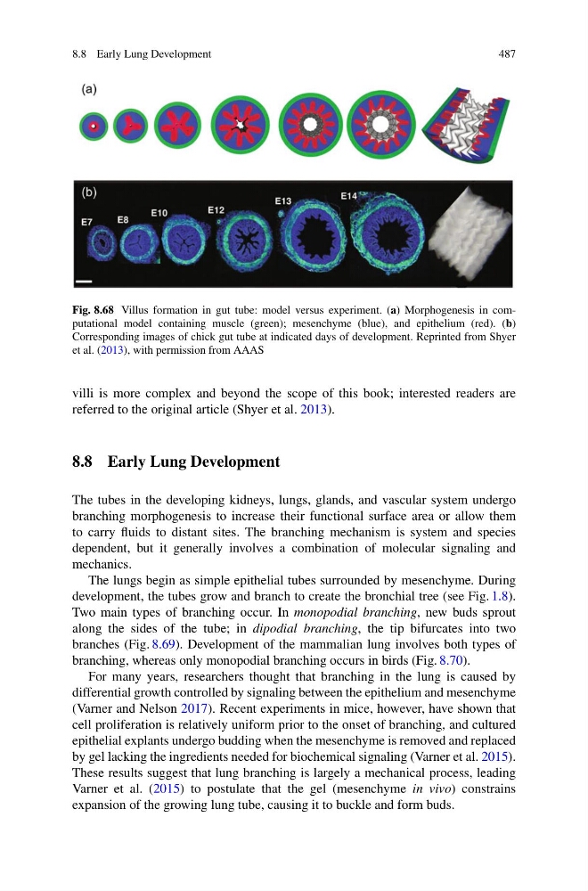
Fig. 8.68 Villus formation in gut tube: model versus experiment. (a) Morphogenesis in computational model containing muscle (green); mesenchyme (blue), and epithelium (red). (b)
Corresponding images of chick gut tube at indicated days of development. Reprinted from Shyer
et al. (2013), with permission from AAAS
villi is more complex and beyond the scope of this book; interested readers are
referred to the original article (Shyer et al. 2013).
8.8 Early Lung Development
The tubes in the developing kidneys, lungs, glands, and vascular system undergo
branching morphogenesis to increase their functional surface area or allow them
to carry fluids to distant sites. The branching mechanism is system and species
dependent, but it generally involves a combination of molecular signaling and
mechanics.
The lungs begin as simple epithelial tubes surrounded by mesenchyme. During
development, the tubes grow and branch to create the bronchial tree (see Fig. 1.8).
Two main types of branching occur. In monopodial branching, new buds sprout
along the sides of the tube; in dipodial branching, the tip bifurcates into two
branches (Fig. 8.69). Development of the mammalian lung involves both types of
branching, whereas only monopodial branching occurs in birds (Fig. 8.70).
For many years, researchers thought that branching in the lung is caused by
differential growth controlled by signaling between the epithelium and mesenchyme
(Varner and Nelson 2017). Recent experiments in mice, however, have shown that
cell proliferation is relatively uniform prior to the onset of branching, and cultured
epithelial explants undergo budding when the mesenchyme is removed and replaced
by gel lacking the ingredients needed for biochemical signaling (Varner et al. 2015).
These results suggest that lung branching is largely a mechanical process, leading
Varner et al. (2015) to postulate that the gel (mesenchyme in vivo) constrains
expansion of the growing lung tube, causing it to buckle and form buds.
487
Fig. 8.68 Villus formation in gut tube: model versus experiment. (a) Morphogenesis in computational model containing muscle (green); mesenchyme (blue), and epithelium (red). (b)
Corresponding images of chick gut tube at indicated days of development. Reprinted from Shyer
et al. (2013), with permission from AAAS
villi is more complex and beyond the scope of this book; interested readers are
referred to the original article (Shyer et al. 2013).
8.8 Early Lung Development
The tubes in the developing kidneys, lungs, glands, and vascular system undergo
branching morphogenesis to increase their functional surface area or allow them
to carry fluids to distant sites. The branching mechanism is system and species
dependent, but it generally involves a combination of molecular signaling and
mechanics.
The lungs begin as simple epithelial tubes surrounded by mesenchyme. During
development, the tubes grow and branch to create the bronchial tree (see Fig. 1.8).
Two main types of branching occur. In monopodial branching, new buds sprout
along the sides of the tube; in dipodial branching, the tip bifurcates into two
branches (Fig. 8.69). Development of the mammalian lung involves both types of
branching, whereas only monopodial branching occurs in birds (Fig. 8.70).
For many years, researchers thought that branching in the lung is caused by
differential growth controlled by signaling between the epithelium and mesenchyme
(Varner and Nelson 2017). Recent experiments in mice, however, have shown that
cell proliferation is relatively uniform prior to the onset of branching, and cultured
epithelial explants undergo budding when the mesenchyme is removed and replaced
by gel lacking the ingredients needed for biochemical signaling (Varner et al. 2015).
These results suggest that lung branching is largely a mechanical process, leading
Varner et al. (2015) to postulate that the gel (mesenchyme in vivo) constrains
expansion of the growing lung tube, causing it to buckle and form buds.