

486
8 Morphogenesis
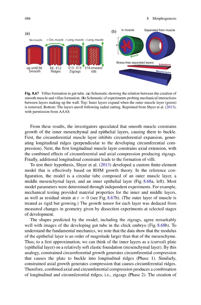
Fig. 8.67 Villus formation in gut tube. (a) Schematic showing the relation between the creation of
smooth muscle and villus formation. (b) Schematic of experiments probing mechanical interactions
between layers making up the wall. Top: Inner layers expand when the outer muscle layer (green)
is removed. Bottom: The layers unroll following radial cutting. Reprinted from Shyer et al. (2013),
with permission from AAAS
From these results, the investigators speculated that smooth muscle constrains
growth of the inner mesenchymal and epithelial layers, causing them to buckle.
First, the circumferential muscle layer inhibits circumferential expansion, generating longitudinal ridges (perpendicular to the developing circumferential compression). Next, the first longitudinal muscle layer constrains axial extension, with
the combined effects of circumferential and axial compression producing zigzags.
Finally, additional longitudinal constraint leads to the formation of villi.
To test their hypothesis, Shyer et al. (2013) developed a custom finite-element
model that is effectively based on RHM growth theory. In the reference configuration, the model is a circular tube composed of an outer muscle layer, a
middle mesenchymal layer, and an inner epithelial layer (Fig. 8.68a, left). Most
model parameters were determined through independent experiments. For example,
mechanical testing provided material properties for the inner and middle layers,
as well as residual strain at t = 0 (see Fig. 8.67b). (The outer layer of muscle is
treated as rigid but growing.) The growth tensor for each layer was deduced from
measured changes in geometry given by dissection experiments at selected stages
of development.
The shapes predicted by the model, including the zigzags, agree remarkably
well with images of the developing gut tube in the chick embryo (Fig. 8.68b). To
understand the fundamental mechanics, we note that the data show that the modulus
of the epithelial layer is an order of magnitude larger than that of the mesenchyme.
Thus, to a first approximation, we can think of the inner layers as a (curved) plate
(epithelial layer) on a relatively soft elastic foundation (mesenchymal layer). By this
analogy, constrained circumferential growth generates circumferential compression
that causes the plate to buckle into longitudinal ridges (Phase 1). Similarly,
constrained axial growth generates compression that causes circumferential ridges.
Therefore, combined axial and circumferential compression produces a combination
of longitudinal and circumferential ridges, i.e., zigzags (Phase 2). The creation of
8 Morphogenesis
Fig. 8.67 Villus formation in gut tube. (a) Schematic showing the relation between the creation of
smooth muscle and villus formation. (b) Schematic of experiments probing mechanical interactions
between layers making up the wall. Top: Inner layers expand when the outer muscle layer (green)
is removed. Bottom: The layers unroll following radial cutting. Reprinted from Shyer et al. (2013),
with permission from AAAS
From these results, the investigators speculated that smooth muscle constrains
growth of the inner mesenchymal and epithelial layers, causing them to buckle.
First, the circumferential muscle layer inhibits circumferential expansion, generating longitudinal ridges (perpendicular to the developing circumferential compression). Next, the first longitudinal muscle layer constrains axial extension, with
the combined effects of circumferential and axial compression producing zigzags.
Finally, additional longitudinal constraint leads to the formation of villi.
To test their hypothesis, Shyer et al. (2013) developed a custom finite-element
model that is effectively based on RHM growth theory. In the reference configuration, the model is a circular tube composed of an outer muscle layer, a
middle mesenchymal layer, and an inner epithelial layer (Fig. 8.68a, left). Most
model parameters were determined through independent experiments. For example,
mechanical testing provided material properties for the inner and middle layers,
as well as residual strain at t = 0 (see Fig. 8.67b). (The outer layer of muscle is
treated as rigid but growing.) The growth tensor for each layer was deduced from
measured changes in geometry given by dissection experiments at selected stages
of development.
The shapes predicted by the model, including the zigzags, agree remarkably
well with images of the developing gut tube in the chick embryo (Fig. 8.68b). To
understand the fundamental mechanics, we note that the data show that the modulus
of the epithelial layer is an order of magnitude larger than that of the mesenchyme.
Thus, to a first approximation, we can think of the inner layers as a (curved) plate
(epithelial layer) on a relatively soft elastic foundation (mesenchymal layer). By this
analogy, constrained circumferential growth generates circumferential compression
that causes the plate to buckle into longitudinal ridges (Phase 1). Similarly,
constrained axial growth generates compression that causes circumferential ridges.
Therefore, combined axial and circumferential compression produces a combination
of longitudinal and circumferential ridges, i.e., zigzags (Phase 2). The creation of