

488
8 Morphogenesis
Fig. 8.69 Two types of
branching in the lung
Dipodial
Monopodial
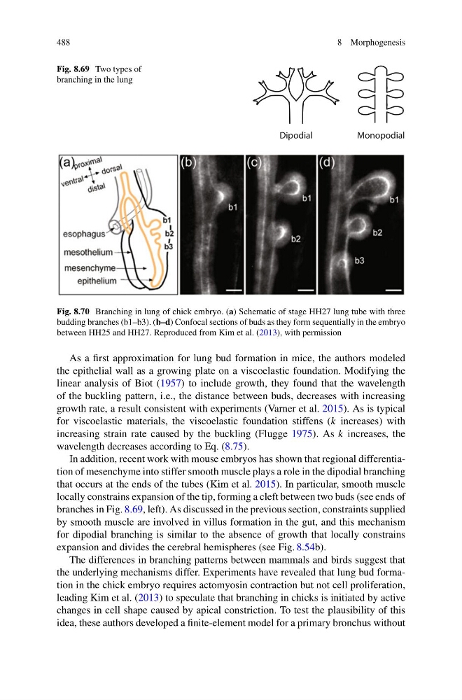
Fig. 8.70 Branching in lung of chick embryo. (a) Schematic of stage HH27 lung tube with three
budding branches (b1–b3). (b–d) Confocal sections of buds as they form sequentially in the embryo
between HH25 and HH27. Reproduced from Kim et al. (2013), with permission
As a first approximation for lung bud formation in mice, the authors modeled
the epithelial wall as a growing plate on a viscoelastic foundation. Modifying the
linear analysis of Biot (1957) to include growth, they found that the wavelength
of the buckling pattern, i.e., the distance between buds, decreases with increasing
growth rate, a result consistent with experiments (Varner et al. 2015). As is typical
for viscoelastic materials, the viscoelastic foundation stiffens (k increases) with
increasing strain rate caused by the buckling (Flugge 1975). As k increases, the
wavelength decreases according to Eq. (8.75).
In addition, recent work with mouse embryos has shown that regional differentiation of mesenchyme into stiffer smooth muscle plays a role in the dipodial branching
that occurs at the ends of the tubes (Kim et al. 2015). In particular, smooth muscle
locally constrains expansion of the tip, forming a cleft between two buds (see ends of
branches in Fig. 8.69, left). As discussed in the previous section, constraints supplied
by smooth muscle are involved in villus formation in the gut, and this mechanism
for dipodial branching is similar to the absence of growth that locally constrains
expansion and divides the cerebral hemispheres (see Fig. 8.54b).
The differences in branching patterns between mammals and birds suggest that
the underlying mechanisms differ. Experiments have revealed that lung bud formation in the chick embryo requires actomyosin contraction but not cell proliferation,
leading Kim et al. (2013) to speculate that branching in chicks is initiated by active
changes in cell shape caused by apical constriction. To test the plausibility of this
idea, these authors developed a finite-element model for a primary bronchus without
8 Morphogenesis
Fig. 8.69 Two types of
branching in the lung
Dipodial
Monopodial
Fig. 8.70 Branching in lung of chick embryo. (a) Schematic of stage HH27 lung tube with three
budding branches (b1–b3). (b–d) Confocal sections of buds as they form sequentially in the embryo
between HH25 and HH27. Reproduced from Kim et al. (2013), with permission
As a first approximation for lung bud formation in mice, the authors modeled
the epithelial wall as a growing plate on a viscoelastic foundation. Modifying the
linear analysis of Biot (1957) to include growth, they found that the wavelength
of the buckling pattern, i.e., the distance between buds, decreases with increasing
growth rate, a result consistent with experiments (Varner et al. 2015). As is typical
for viscoelastic materials, the viscoelastic foundation stiffens (k increases) with
increasing strain rate caused by the buckling (Flugge 1975). As k increases, the
wavelength decreases according to Eq. (8.75).
In addition, recent work with mouse embryos has shown that regional differentiation of mesenchyme into stiffer smooth muscle plays a role in the dipodial branching
that occurs at the ends of the tubes (Kim et al. 2015). In particular, smooth muscle
locally constrains expansion of the tip, forming a cleft between two buds (see ends of
branches in Fig. 8.69, left). As discussed in the previous section, constraints supplied
by smooth muscle are involved in villus formation in the gut, and this mechanism
for dipodial branching is similar to the absence of growth that locally constrains
expansion and divides the cerebral hemispheres (see Fig. 8.54b).
The differences in branching patterns between mammals and birds suggest that
the underlying mechanisms differ. Experiments have revealed that lung bud formation in the chick embryo requires actomyosin contraction but not cell proliferation,
leading Kim et al. (2013) to speculate that branching in chicks is initiated by active
changes in cell shape caused by apical constriction. To test the plausibility of this
idea, these authors developed a finite-element model for a primary bronchus without