

8.5 Early Heart Development
453
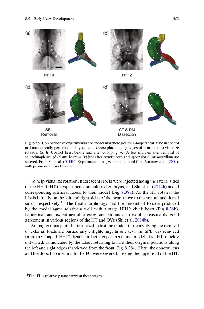
Fig. 8.38 Comparison of experimental and model morphologies for c-looped heart tube in control
and mechanically perturbed embryos. Labels were placed along edges of heart tube to visualize
rotation. (a, b) Control heart before and after c-looping. (c) A few minutes after removal of
splanchnopleure. (d) Same heart as (c) just after conotruncus and upper dorsal mesocardium are
severed. From Shi et al. (2014b). Experimental images are reproduced from Voronov et al. (2004),
with permission from Elsevier
To help visualize rotation, fluorescent labels were injected along the lateral sides
of the HH10 HT in experiments on cultured embryos, and Shi et al. (2014b) added
corresponding artificial labels to their model (Fig. 8.38a). As the HT rotates, the
labels initially on the left and right sides of the heart move to the ventral and dorsal
sides, respectively. 11 The final morphology and the amount of torsion produced
by the model agree relatively well with a stage HH12 chick heart (Fig. 8.38b).
Numerical and experimental stresses and strains also exhibit reasonably good
agreement in various regions of the HT and OVs (Shi et al. 2014b).
Among various perturbations used to test the model, those involving the removal
of external loads are particularly enlightening. In one test, the SPL was removed
from the looped HH12 heart. In both experiment and model, the HT quickly
untwisted, as indicated by the labels returning toward their original positions along
the left and right edges (as viewed from the front; Fig. 8.38c). Next, the conotruncus
and the dorsal connection to the FG were severed, freeing the upper end of the HT.
11 The HT is relatively transparent at these stages.
453
Fig. 8.38 Comparison of experimental and model morphologies for c-looped heart tube in control
and mechanically perturbed embryos. Labels were placed along edges of heart tube to visualize
rotation. (a, b) Control heart before and after c-looping. (c) A few minutes after removal of
splanchnopleure. (d) Same heart as (c) just after conotruncus and upper dorsal mesocardium are
severed. From Shi et al. (2014b). Experimental images are reproduced from Voronov et al. (2004),
with permission from Elsevier
To help visualize rotation, fluorescent labels were injected along the lateral sides
of the HH10 HT in experiments on cultured embryos, and Shi et al. (2014b) added
corresponding artificial labels to their model (Fig. 8.38a). As the HT rotates, the
labels initially on the left and right sides of the heart move to the ventral and dorsal
sides, respectively. 11 The final morphology and the amount of torsion produced
by the model agree relatively well with a stage HH12 chick heart (Fig. 8.38b).
Numerical and experimental stresses and strains also exhibit reasonably good
agreement in various regions of the HT and OVs (Shi et al. 2014b).
Among various perturbations used to test the model, those involving the removal
of external loads are particularly enlightening. In one test, the SPL was removed
from the looped HH12 heart. In both experiment and model, the HT quickly
untwisted, as indicated by the labels returning toward their original positions along
the left and right edges (as viewed from the front; Fig. 8.38c). Next, the conotruncus
and the dorsal connection to the FG were severed, freeing the upper end of the HT.
11 The HT is relatively transparent at these stages.