

452
8 Morphogenesis
CJ
CJ
Lumen
CJ
CJ
MY
SPL
AIP
FG
Ventral
Cranial
Left
CT
HT
OV
OV
MY
CJ
Lumen
SPL
FG
DM
200 m
P
Lumen
Ventral
Left
DM
}
}
Cranial
Ventral
Left
AIP
ROV
LOV
HT
CT
Endodermal Contraction
Isotropic Growth
MY Differential Growth,
MY Cell-Shape Change,
& OV Fusion
CJ Growth & OV Fusion
DM Tension
)
c
(
)
b
(
)
a
(
)
’
b
(
)
’
a
(
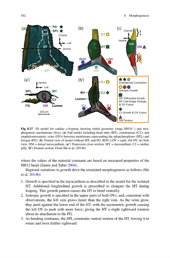
Fig. 8.37 3D model for cardiac c-looping showing initial geometry (stage HH10 − ) and morphogenetic mechanisms (box). (a) Full model including heart tube (HT), conotruncus (CT), and
omphalomesenteric veins (OVs) between membranes representing the splanchnopleure (SPL) and
foregut (FG). (b) Ventral view of model without SPL and FG. ROV, LOV = right, left OV. (c) Side
view. DM = dorsal mesocardium. (a’) Transverse cross section. MY = myocardium; CJ = cardiac
jelly. (b’) Frontal section. From Shi et al. (2014b)
where the values of the material constants are based on measured properties of the
HH12 heart (Zamir and Taber 2004).
Regional variations in growth drive the simulated morphogenesis as follows (Shi
et al. 2014b):
1. Growth is specified in the myocardium as described in the model for the isolated
HT. Additional longitudinal growth is prescribed to elongate the HT during
looping. This growth pattern causes the HT to bend ventrally.
2. Isotropic growth is specified in the upper parts of both OVs, and, consistent with
observations, the left vein grows faster than the right vein. As the veins grow,
they push against the lower end of the HT, with the asymmetric growth causing
the left OV to push with more force, giving the HT a slight rightward rotation
about its attachment to the FG.
3. As bending continues, the SPL constrains ventral motion of the HT, forcing it to
rotate and twist further rightward.
8 Morphogenesis
CJ
CJ
Lumen
CJ
CJ
MY
SPL
AIP
FG
Ventral
Cranial
Left
CT
HT
OV
OV
MY
CJ
Lumen
SPL
FG
DM
200 m
P
Lumen
Ventral
Left
DM
}
}
Cranial
Ventral
Left
AIP
ROV
LOV
HT
CT
Endodermal Contraction
Isotropic Growth
MY Differential Growth,
MY Cell-Shape Change,
& OV Fusion
CJ Growth & OV Fusion
DM Tension
)
c
(
)
b
(
)
a
(
)
’
b
(
)
’
a
(
Fig. 8.37 3D model for cardiac c-looping showing initial geometry (stage HH10 − ) and morphogenetic mechanisms (box). (a) Full model including heart tube (HT), conotruncus (CT), and
omphalomesenteric veins (OVs) between membranes representing the splanchnopleure (SPL) and
foregut (FG). (b) Ventral view of model without SPL and FG. ROV, LOV = right, left OV. (c) Side
view. DM = dorsal mesocardium. (a’) Transverse cross section. MY = myocardium; CJ = cardiac
jelly. (b’) Frontal section. From Shi et al. (2014b)
where the values of the material constants are based on measured properties of the
HH12 heart (Zamir and Taber 2004).
Regional variations in growth drive the simulated morphogenesis as follows (Shi
et al. 2014b):
1. Growth is specified in the myocardium as described in the model for the isolated
HT. Additional longitudinal growth is prescribed to elongate the HT during
looping. This growth pattern causes the HT to bend ventrally.
2. Isotropic growth is specified in the upper parts of both OVs, and, consistent with
observations, the left vein grows faster than the right vein. As the veins grow,
they push against the lower end of the HT, with the asymmetric growth causing
the left OV to push with more force, giving the HT a slight rightward rotation
about its attachment to the FG.
3. As bending continues, the SPL constrains ventral motion of the HT, forcing it to
rotate and twist further rightward.