

8.5 Early Heart Development
451
The quantitative differences between model and experiment could indicate that
something about the model is not realistic (besides the idealized geometry). Perhaps
one or more of the assumptions are wrong. However, the fact that the model
captures the correct trends in the data is not a trivial result. For example, the model
correctly predicts that the initial tension decreases in both directions throughout
the myocardium as the HT bends. In contrast, bending by externally applied loads
would compress the tube axially at the inner curvature and stretch it at the outer
curvature, causing the longitudinal tension to increase on the convex side. Therefore,
the model generally supports the notion that differential growth is the primary driver
of the bending component of cardiac c-looping, with smaller contributions from
other mechanisms, such as active changes in myocardial cell shape.
Twisting the Heart Tube Abnormal looping is associated with many of the
congenital heart defects that occur in about 1% of live human births (Hoffman
1995). The most conspicuous abnormality is the relatively rare instance where the
heart loops to the left rather than to the right. If the heart is not oriented properly,
the major blood vessels may not connect in the right locations, leading to severe
consequences.
The torsional component of looping determines left-right directionality. Much
remains unknown about this process. To minimize defects, however, it is likely
that multiple mechanisms are involved in steering the HT in the “right” direction,
i.e., toward the right. Some speculate that these processes generate relatively small
asymmetries that bias rightward rotation, with external loads driving most of the
ensuing torsional deformation (Voronov et al. 2004; Desgrange et al. 2018). The
initial steering mechanisms could include asymmetries in the shape of the HT and
the sizes of the OVs prior to looping (Voronov et al. 2004; Kidokoro et al. 2008),
differences in cell proliferation on the right and left sides of the dorsal mesocardium
(Linask et al. 2005), asymmetric contraction in the HT or AIP (Itasaki et al. 1991;
Shi et al. 2015), and intrinsic rotation of myocardial cells before and during looping
(Ray et al. 2018).
Recently, Shi et al. (2014b) presented a 3D finite-element model for c-looping
that includes both bending and torsion. The initial geometry was constructed from
cross-sectional images of a stage HH10 − chick heart. The model includes the
HT, OVs, and conotruncus (outflow tract), all sandwiched between two frictionless
membranes representing the SPL and ventral wall of the FG (Fig. 8.37). The ends
of the OVs are constrained to move only laterally, and the dorsal mesocardium (the
dorsal side of the HT) is assumed to be free (ruptured) except at its upper and lower
ends, which are connected by vertical rollers to the FG. The wall of the model HT
consists of myocardium and cardiac jelly (Fig. 8.37a’, b’). The strain-energy density
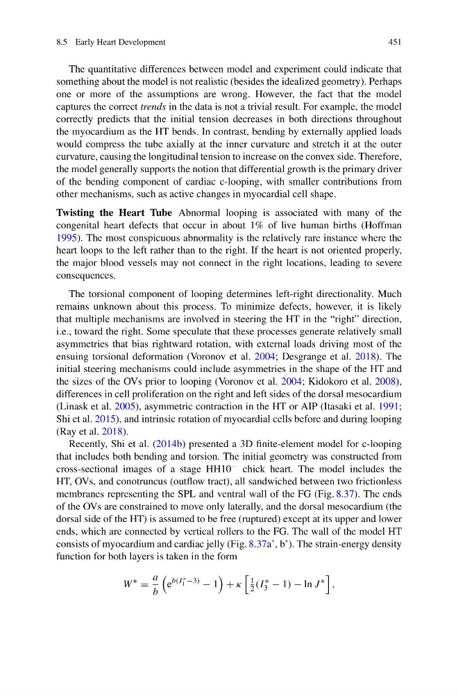
function for both layers is taken in the form
W
∗
=
a
b
e
b(I ∗
1 −3)
− 1
+ κ
1
2 (I
∗
3 − 1) − ln J
∗
,
451
The quantitative differences between model and experiment could indicate that
something about the model is not realistic (besides the idealized geometry). Perhaps
one or more of the assumptions are wrong. However, the fact that the model
captures the correct trends in the data is not a trivial result. For example, the model
correctly predicts that the initial tension decreases in both directions throughout
the myocardium as the HT bends. In contrast, bending by externally applied loads
would compress the tube axially at the inner curvature and stretch it at the outer
curvature, causing the longitudinal tension to increase on the convex side. Therefore,
the model generally supports the notion that differential growth is the primary driver
of the bending component of cardiac c-looping, with smaller contributions from
other mechanisms, such as active changes in myocardial cell shape.
Twisting the Heart Tube Abnormal looping is associated with many of the
congenital heart defects that occur in about 1% of live human births (Hoffman
1995). The most conspicuous abnormality is the relatively rare instance where the
heart loops to the left rather than to the right. If the heart is not oriented properly,
the major blood vessels may not connect in the right locations, leading to severe
consequences.
The torsional component of looping determines left-right directionality. Much
remains unknown about this process. To minimize defects, however, it is likely
that multiple mechanisms are involved in steering the HT in the “right” direction,
i.e., toward the right. Some speculate that these processes generate relatively small
asymmetries that bias rightward rotation, with external loads driving most of the
ensuing torsional deformation (Voronov et al. 2004; Desgrange et al. 2018). The
initial steering mechanisms could include asymmetries in the shape of the HT and
the sizes of the OVs prior to looping (Voronov et al. 2004; Kidokoro et al. 2008),
differences in cell proliferation on the right and left sides of the dorsal mesocardium
(Linask et al. 2005), asymmetric contraction in the HT or AIP (Itasaki et al. 1991;
Shi et al. 2015), and intrinsic rotation of myocardial cells before and during looping
(Ray et al. 2018).
Recently, Shi et al. (2014b) presented a 3D finite-element model for c-looping
that includes both bending and torsion. The initial geometry was constructed from
cross-sectional images of a stage HH10 − chick heart. The model includes the
HT, OVs, and conotruncus (outflow tract), all sandwiched between two frictionless
membranes representing the SPL and ventral wall of the FG (Fig. 8.37). The ends
of the OVs are constrained to move only laterally, and the dorsal mesocardium (the
dorsal side of the HT) is assumed to be free (ruptured) except at its upper and lower
ends, which are connected by vertical rollers to the FG. The wall of the model HT
consists of myocardium and cardiac jelly (Fig. 8.37a’, b’). The strain-energy density
function for both layers is taken in the form
W
∗
=
a
b
e
b(I ∗
1 −3)
− 1
+ κ
1
2 (I
∗
3 − 1) − ln J
∗
,