

vesicles which fuse with the plasma membrane in the distal portion of the cell,
transferring the secretion to the periplasmic space (Fig. 11) [139, 144, 145, 147,
151]. Then, the secretion crosses the cell wall and cuticle, reaching the gland surface
and escaping to the atmosphere. Diffusion of the secretion across the cuticle can
occur due to the lipophilic nature of cutin, aided by the secretory flow, the pressure
exerted by the protoplast, and/or the possible presence of microchannels in the
cuticle [23].
3.2
The Chemical Nature of the Odor
The floral odors produced by osmophores are usually composed of isoprenoids,
terpenoids, benzenoids, phenylpropanoids, fatty acid derivatives, and various
nitrogenous and sulfur-containing compounds. These are generally of low molecular
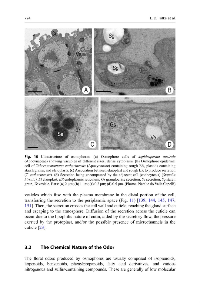
Fig. 10 Ultrastructure of osmophores. (a) Osmophore cells of Aspidosperma australe
(Apocynaceae) showing vacuoles of different sizes; dense cytoplasm. (b) Osmophore epidermal
cell of Tabernaemontana catharinensis (Apocynaceae) containing rough ER, plastids containing
starch grains, and elaioplasts. (c) Association between elaioplast and rough ER to produce secretion
(T. catharinensis). (d) Secretion being encompassed by the adjacent cell (endocytosis) (Stapelia
hirsuta). El elaioplast, ER endoplasmic reticulum, Gs granulocrine secretion, Se secretion, Sg starch
grain, Ve vesicle. Bars: (a) 2 μm; (b) 1 μm; (c) 0.2 μm; (d) 0.5 μm. (Photos: Natalie do Valle Capelli)
724
E. D. Tölke et al.