

3.1
Osmophore Structure, Odor Production, and Release
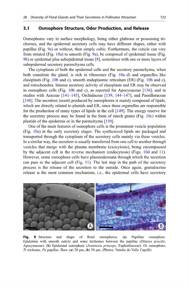
Osmophores vary in surface morphology, being either glabrous or possessing trichomes, and the epidermal secretory cells may have different shapes, either with
papillae (Fig. 9a) or without, then simply cubic. Furthermore, the cuticle can vary
from striated (Fig. 10a) to smooth (Fig. 9a), be composed of epidermal tissue (Fig.
9b) or epidermal plus subepidermal tissue [8], sometimes with one or more layers of
subepidermal secretory parenchyma cells.
The cytoplasm of both the epidermal cells and the secretory parenchyma, when
both constitute the gland, is rich in ribosomes (Fig. 10a–d) and organelles like
elaioplasts (Fig. 10b and c), smooth endoplasmic reticulum (ER) (Fig. 10b and c),
and mitochondria. Intense secretory activity of elaioplasts and ER may be observed
in osmophore cells (Fig. 10b and c), as reported for Apocynaceae [136], and in
studies with Araceae [141–143], Orchidaceae [139, 144–147], and Passifloraceae
[148]. The secretion (scent) produced by osmophores is mainly composed of lipids,
which are directly related to plastids and ER, since these organelles are responsible
for the production of many types of lipids in the cell [149]. The energy reserve for
the secretory process may be found in the form of starch grains (Fig. 10c) within
plastids of the epidermis or in the parenchyma [150].
One of the main features of osmophore cells is the prominent vesicle population
(Fig. 10a) in the early secretory stages. The synthesized lipids are packaged and
transported through the cytoplasm of the secretory cells mainly via these vesicles.
In a similar way, the secretion is usually transferred from one cell to another through
vesicles that merge with the plasma membrane (exocytosis), being encompassed
by the adjacent cell in the reverse mechanism (endocytosis) (Figs. 10d and 11).
However, some osmophore cells have plasmodesmata through which the secretion
can pass to the adjacent cell (Fig. 11). The last step in the path of the secretory
process is the release of the secretion to the outside. Once again, granulocrine
release is the most common mechanism, i.e., the epidermal cells have secretory
Fig. 9 Structure and shape of floral osmophores. (a) Papillate osmophore.
Epidermis with smooth cuticle and some trichomes between the papillae (Ditassa gracilis,
Apocynaceae). (b) Epidermal osmophore (Joannesia princeps, Euphorbiaceae). Os osmophore,
Tr trichome, Pa papillae. Bars: (a) 20 μm, (b) 50 μm. (Photos: Natalie do Valle Capelli)
28 Diversity of Floral Glands and Their Secretions in Pollinator Attraction
723