

and less dense cytoplasm [10, 11, 14–16]. Vascular bundles are usually present in
the subnectary parenchyma (Fig. 1a and b) and may contain phloem or xylem or
both [14–16]. The vascular supply in some cases is correlated with the sugar
concentration in the nectar. Nectaries that secrete very concentrated nectar are
vascularized by phloem only, while low sugar concentrations occur in nectaries
vascularized equally by xylem and phloem or only by xylem [25, 37, 47–49]. The
vascular branches may reach the nectary parenchyma and sometimes even
the epidermis, but generally just the phloem elements are found among these tissues
[16, 44, 50].
The ultrastructure of nectariferous tissues is quite similar in species from different
lineages of plants and have numerous vacuoles that increase their size along
the secretory process, dense cytoplasm rich in ribosomes, highly developed rough
endoplasmic reticulum, active dictyosomes, numerous mitochondria, and plastids
sometimes containing large starch granules (Fig. 3a–d) [10, 11, 15]. The vacuoles
are small and numerous in early phases of the secretory process in the course of which
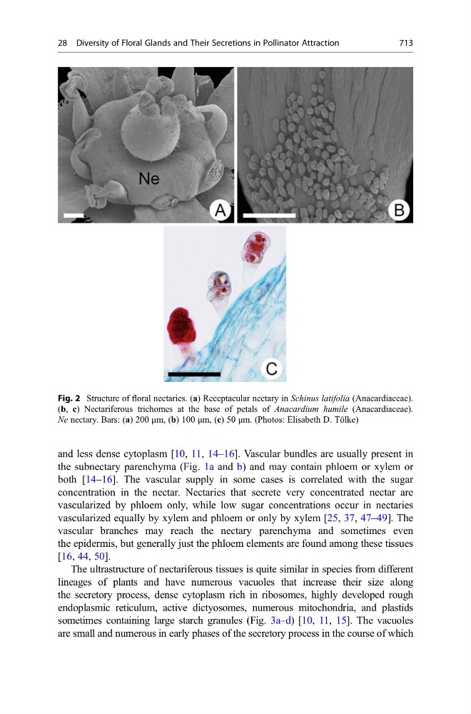
Fig. 2 Structure of floral nectaries. (a) Receptacular nectary in Schinus latifolia (Anacardiaceae).
(b, c) Nectariferous trichomes at the base of petals of Anacardium humile (Anacardiaceae).
Ne nectary. Bars: (a) 200 μm, (b) 100 μm, (c) 50 μm. (Photos: Elisabeth D. Tölke)
28 Diversity of Floral Glands and Their Secretions in Pollinator Attraction
713