

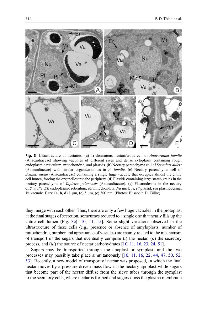
they merge with each other. Thus, there are only a few huge vacuoles in the protoplast
at the final stages of secretion, sometimes reduced to a single one that nearly fills up the
entire cell lumen (Fig. 3c) [10, 11, 15]. Some slight variations observed in the
ultrastructure of these cells (e.g., presence or absence of amyloplasts, number of
mitochondria, number and appearance of vesicles) are mainly related to the mechanism
of transport of the sugars that eventually compose (i) the nectar, (ii) the secretory
process, and (iii) the source of nectar carbohydrates [10, 11, 16, 23, 24, 51].
Sugars may be transported through the apoplast or symplast, and the two
processes may possibly take place simultaneously [10, 11, 16, 22, 44, 47, 50, 52,
53]. Recently, a new model of transport of nectar was proposed, in which the final
nectar moves by a pressure-driven mass flow in the nectary apoplast while sugars
that become part of the nectar diffuse from the sieve tubes through the symplast
to the secretory cells, where nectar is formed and sugars cross the plasma membrane
Fig. 3 Ultrastructure of nectaries. (a) Trichomatous nectariferous cell of Anacardium humile
(Anacardiaceae) showing vacuoles of different sizes and dense cytoplasm containing rough
endoplasmic reticulum, mitochondria, and plastids. (b) Nectary parenchyma cell of Spondias dulcis
(Anacardiaceae) with similar organization as in A. humile. (c) Nectary parenchyma cell of
Schinus molle (Anacardiaceae) containing a single huge vacuole that occupies almost the entire
cell lumen, forcing the organelles into the periphery. (d) Plastids containing large starch grains in the
nectary parenchyma of Tapirira guianensis (Anacardiaceae). (e) Plasmodesma in the nectary
of S. molle. ER endoplasmic reticulum, Mi mitochondria, Nu nucleus, Pl plastid, Pm plasmodesma,
Va vacuole. Bars: (a, b, d) 1 μm, (c) 5 μm, (e) 500 nm. (Photos: Elisabeth D. Tölke)
714
E. D. Tölke et al.