

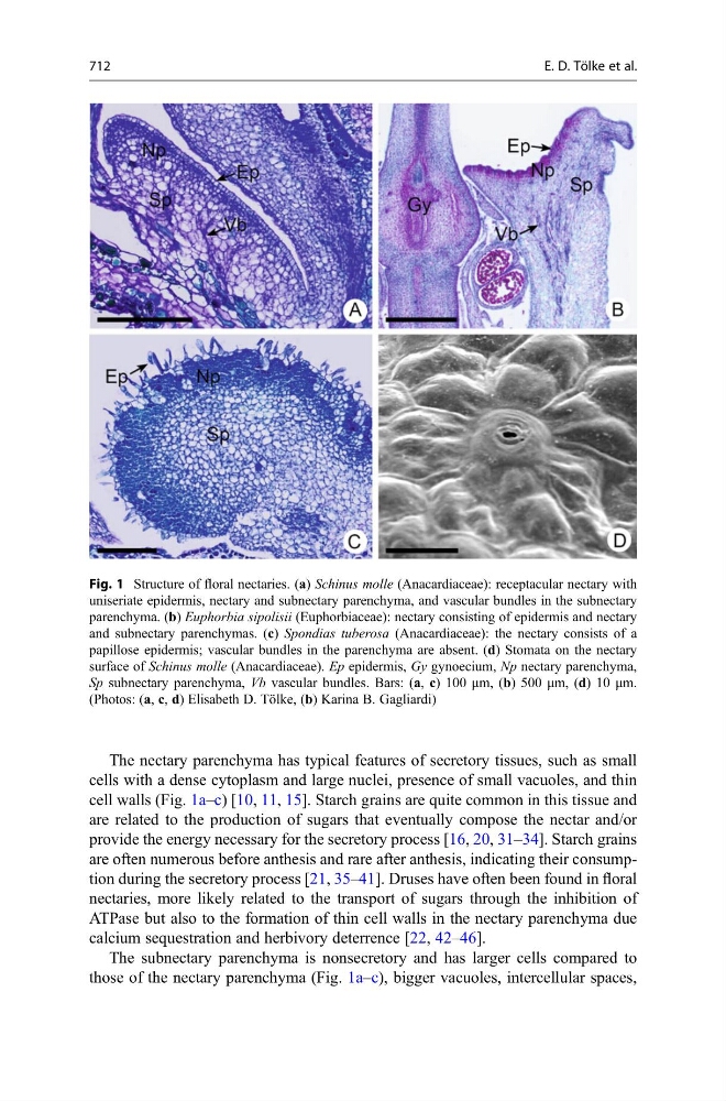
The nectary parenchyma has typical features of secretory tissues, such as small
cells with a dense cytoplasm and large nuclei, presence of small vacuoles, and thin
cell walls (Fig. 1a–c) [10, 11, 15]. Starch grains are quite common in this tissue and
are related to the production of sugars that eventually compose the nectar and/or
provide the energy necessary for the secretory process [16, 20, 31–34]. Starch grains
are often numerous before anthesis and rare after anthesis, indicating their consumption during the secretory process [21, 35–41]. Druses have often been found in floral
nectaries, more likely related to the transport of sugars through the inhibition of
ATPase but also to the formation of thin cell walls in the nectary parenchyma due
calcium sequestration and herbivory deterrence [22, 42–46].
The subnectary parenchyma is nonsecretory and has larger cells compared to
those of the nectary parenchyma (Fig. 1a–c), bigger vacuoles, intercellular spaces,
Fig. 1 Structure of floral nectaries. (a) Schinus molle (Anacardiaceae): receptacular nectary with
uniseriate epidermis, nectary and subnectary parenchyma, and vascular bundles in the subnectary
parenchyma. (b) Euphorbia sipolisii (Euphorbiaceae): nectary consisting of epidermis and nectary
and subnectary parenchymas. (c) Spondias tuberosa (Anacardiaceae): the nectary consists of a
papillose epidermis; vascular bundles in the parenchyma are absent. (d) Stomata on the nectary
surface of Schinus molle (Anacardiaceae). Ep epidermis, Gy gynoecium, Np nectary parenchyma,
Sp subnectary parenchyma, Vb vascular bundles. Bars: (a, c) 100 μm, (b) 500 μm, (d) 10 μm.
(Photos: (a, c, d) Elisabeth D. Tölke, (b) Karina B. Gagliardi)
712
E. D. Tölke et al.