

282
Effect on Lipid Metabolism
Several evidences are available to indicate that exposure of MDCs during developing stage can induce severe metabolic disorders (Fig. 17.9) such as obesity, insulin
resistance, hormonal imbalance, and hyperlipidemia [75]. Adipogenesis regulating
genes such as PPARɤ or some other related genes are main targets for EDCs. During
the developing stage, differentiation of mesenchymal stem cells into adipocytes is
regulated by PPARɤ. In vitro study has indicated that when cell lines of mouse
embryo exposed to tributyltin, it caused the decrease in DNA methylation in promoter region by increasing the differentiation into adipocytes [76]. Such adipogenesis was also observed in mesenchymal stem cells of human when exposed to
dibutyltin (DBT), a metabolite of tributyltin. More precisely, the mesenchymal cells
of human are more receptive to the treatment than MSC mouse, with C/EBPα and
PPARγ2 that regulate each other with a positive feedback loop and act as significant
indicators for adipose differentiation. Over expression of fat-specific protein-27
(FSP27), (FSP4), and LPL were also seen in these cells leading to fatty liver diseases [77]. When sucrose rich fat diet was given to Sprague-Dawley rats then
non- phenolic co-administration (NP) increased the food and water intake in mouse
along with hepatic echogenicity with changes in many plasmatic aminotransferases.
The hepatic echogenicity causes elasticity problems in central vein, inflammatory
cell infiltration, and up-regulation of genes inducing lipogenesis such as srebp-1C,
fas, and ucp2 [78].
Exposure of triclosan (TCS) to human mesenchymal cells can cause lipid accumulation by decreasing the expression of adipocyte protein 2 (ap2), lpl, and adiponectin (adipoq) gene [79]. Oral exposure of DEHP to rats with dose amount of 0.05,
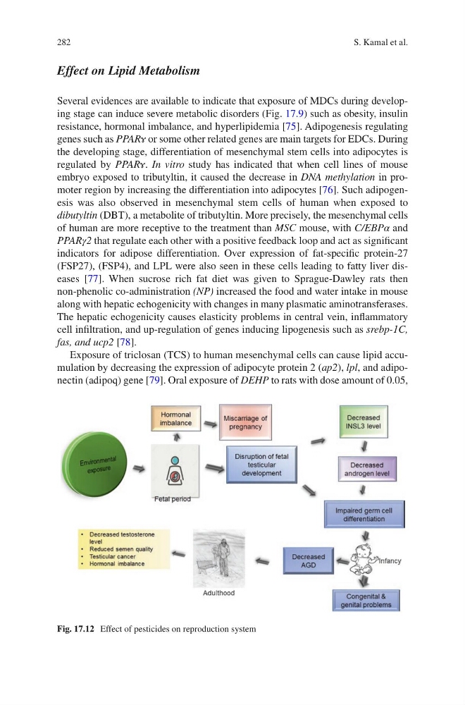
Fig. 17.12 Effect of pesticides on reproduction system
S. Kamal et al.
Effect on Lipid Metabolism
Several evidences are available to indicate that exposure of MDCs during developing stage can induce severe metabolic disorders (Fig. 17.9) such as obesity, insulin
resistance, hormonal imbalance, and hyperlipidemia [75]. Adipogenesis regulating
genes such as PPARɤ or some other related genes are main targets for EDCs. During
the developing stage, differentiation of mesenchymal stem cells into adipocytes is
regulated by PPARɤ. In vitro study has indicated that when cell lines of mouse
embryo exposed to tributyltin, it caused the decrease in DNA methylation in promoter region by increasing the differentiation into adipocytes [76]. Such adipogenesis was also observed in mesenchymal stem cells of human when exposed to
dibutyltin (DBT), a metabolite of tributyltin. More precisely, the mesenchymal cells
of human are more receptive to the treatment than MSC mouse, with C/EBPα and
PPARγ2 that regulate each other with a positive feedback loop and act as significant
indicators for adipose differentiation. Over expression of fat-specific protein-27
(FSP27), (FSP4), and LPL were also seen in these cells leading to fatty liver diseases [77]. When sucrose rich fat diet was given to Sprague-Dawley rats then
non- phenolic co-administration (NP) increased the food and water intake in mouse
along with hepatic echogenicity with changes in many plasmatic aminotransferases.
The hepatic echogenicity causes elasticity problems in central vein, inflammatory
cell infiltration, and up-regulation of genes inducing lipogenesis such as srebp-1C,
fas, and ucp2 [78].
Exposure of triclosan (TCS) to human mesenchymal cells can cause lipid accumulation by decreasing the expression of adipocyte protein 2 (ap2), lpl, and adiponectin (adipoq) gene [79]. Oral exposure of DEHP to rats with dose amount of 0.05,
Fig. 17.12 Effect of pesticides on reproduction system
S. Kamal et al.