

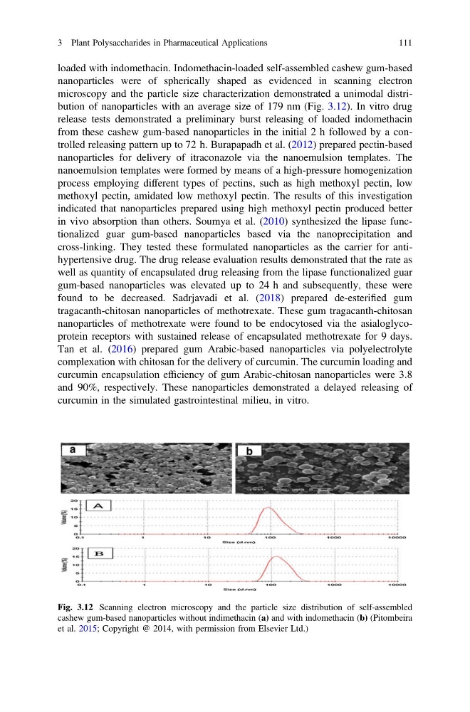
loaded with indomethacin. Indomethacin-loaded self-assembled cashew gum-based
nanoparticles were of spherically shaped as evidenced in scanning electron
microscopy and the particle size characterization demonstrated a unimodal distribution of nanoparticles with an average size of 179 nm (Fig. 3.12). In vitro drug
release tests demonstrated a preliminary burst releasing of loaded indomethacin
from these cashew gum-based nanoparticles in the initial 2 h followed by a controlled releasing pattern up to 72 h. Burapapadh et al. (2012) prepared pectin-based
nanoparticles for delivery of itraconazole via the nanoemulsion templates. The
nanoemulsion templates were formed by means of a high-pressure homogenization
process employing different types of pectins, such as high methoxyl pectin, low
methoxyl pectin, amidated low methoxyl pectin. The results of this investigation
indicated that nanoparticles prepared using high methoxyl pectin produced better
in vivo absorption than others. Soumya et al. (2010) synthesized the lipase functionalized guar gum-based nanoparticles based via the nanoprecipitation and
cross-linking. They tested these formulated nanoparticles as the carrier for antihypertensive drug. The drug release evaluation results demonstrated that the rate as
well as quantity of encapsulated drug releasing from the lipase functionalized guar
gum-based nanoparticles was elevated up to 24 h and subsequently, these were
found to be decreased. Sadrjavadi et al. (2018) prepared de-esterified gum
tragacanth-chitosan nanoparticles of methotrexate. These gum tragacanth-chitosan
nanoparticles of methotrexate were found to be endocytosed via the asialoglycoprotein receptors with sustained release of encapsulated methotrexate for 9 days.
Tan et al. (2016) prepared gum Arabic-based nanoparticles via polyelectrolyte
complexation with chitosan for the delivery of curcumin. The curcumin loading and
curcumin encapsulation efficiency of gum Arabic-chitosan nanoparticles were 3.8
and 90%, respectively. These nanoparticles demonstrated a delayed releasing of
curcumin in the simulated gastrointestinal milieu, in vitro.
Fig. 3.12 Scanning electron microscopy and the particle size distribution of self-assembled
cashew gum-based nanoparticles without indimethacin (a) and with indomethacin (b) (Pitombeira
et al. 2015; Copyright @ 2014, with permission from Elsevier Ltd.)
3 Plant Polysaccharides in Pharmaceutical Applications
111
nanoparticles were of spherically shaped as evidenced in scanning electron
microscopy and the particle size characterization demonstrated a unimodal distribution of nanoparticles with an average size of 179 nm (Fig. 3.12). In vitro drug
release tests demonstrated a preliminary burst releasing of loaded indomethacin
from these cashew gum-based nanoparticles in the initial 2 h followed by a controlled releasing pattern up to 72 h. Burapapadh et al. (2012) prepared pectin-based
nanoparticles for delivery of itraconazole via the nanoemulsion templates. The
nanoemulsion templates were formed by means of a high-pressure homogenization
process employing different types of pectins, such as high methoxyl pectin, low
methoxyl pectin, amidated low methoxyl pectin. The results of this investigation
indicated that nanoparticles prepared using high methoxyl pectin produced better
in vivo absorption than others. Soumya et al. (2010) synthesized the lipase functionalized guar gum-based nanoparticles based via the nanoprecipitation and
cross-linking. They tested these formulated nanoparticles as the carrier for antihypertensive drug. The drug release evaluation results demonstrated that the rate as
well as quantity of encapsulated drug releasing from the lipase functionalized guar
gum-based nanoparticles was elevated up to 24 h and subsequently, these were
found to be decreased. Sadrjavadi et al. (2018) prepared de-esterified gum
tragacanth-chitosan nanoparticles of methotrexate. These gum tragacanth-chitosan
nanoparticles of methotrexate were found to be endocytosed via the asialoglycoprotein receptors with sustained release of encapsulated methotrexate for 9 days.
Tan et al. (2016) prepared gum Arabic-based nanoparticles via polyelectrolyte
complexation with chitosan for the delivery of curcumin. The curcumin loading and
curcumin encapsulation efficiency of gum Arabic-chitosan nanoparticles were 3.8
and 90%, respectively. These nanoparticles demonstrated a delayed releasing of
curcumin in the simulated gastrointestinal milieu, in vitro.
Fig. 3.12 Scanning electron microscopy and the particle size distribution of self-assembled
cashew gum-based nanoparticles without indimethacin (a) and with indomethacin (b) (Pitombeira
et al. 2015; Copyright @ 2014, with permission from Elsevier Ltd.)
3 Plant Polysaccharides in Pharmaceutical Applications
111