

62
S. Kakkar et al.
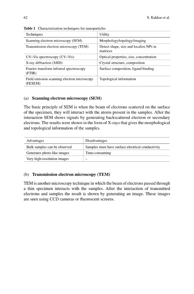
Table 1 Characterization techniques for nanoparticles
Techniques
Utility
Scanning electron microscopy (SEM)
Morphology/topology/imaging
Transmission electron microscopy (TEM)
Detect shape, size and localize NPs in
matrices
UV–Vis spectroscopy (UV–Vis)
Optical properties, size, concentration
X-ray diffraction (XRD)
Crystal structure, composition
Fourier transform infrared spectroscopy
(FTIR)
Surface composition, ligand binding
Field emission scanning electron microscopy
(FESEM)
Topological information
(a) Scanning electron microscopy (SEM)
The basic principle of SEM is when the beam of electrons scattered on the surface
of the specimen, they will interact with the atoms present in the samples. After the
interaction SEM shows signals by generating backscattered electron or secondary
electrons. The results were shown in the form of X-rays that gives the morphological
and topological information of the samples.
Advantages
Disadvantages
Bulk samples can be observed
Samples must have surface electrical conductivity
Generates photo-like images
Time-consuming
Very high-resolution images
–
(b) Transmission electron microscopy (TEM)
TEM is another microscopy technique in which the beam of electrons passed through
a thin specimen interacts with the samples. After the interaction of transmitted
electrons and samples the result is shown by generating an image. These images
are seen using CCD cameras or fluorescent screens.
S. Kakkar et al.
Table 1 Characterization techniques for nanoparticles
Techniques
Utility
Scanning electron microscopy (SEM)
Morphology/topology/imaging
Transmission electron microscopy (TEM)
Detect shape, size and localize NPs in
matrices
UV–Vis spectroscopy (UV–Vis)
Optical properties, size, concentration
X-ray diffraction (XRD)
Crystal structure, composition
Fourier transform infrared spectroscopy
(FTIR)
Surface composition, ligand binding
Field emission scanning electron microscopy
(FESEM)
Topological information
(a) Scanning electron microscopy (SEM)
The basic principle of SEM is when the beam of electrons scattered on the surface
of the specimen, they will interact with the atoms present in the samples. After the
interaction SEM shows signals by generating backscattered electron or secondary
electrons. The results were shown in the form of X-rays that gives the morphological
and topological information of the samples.
Advantages
Disadvantages
Bulk samples can be observed
Samples must have surface electrical conductivity
Generates photo-like images
Time-consuming
Very high-resolution images
–
(b) Transmission electron microscopy (TEM)
TEM is another microscopy technique in which the beam of electrons passed through
a thin specimen interacts with the samples. After the interaction of transmitted
electrons and samples the result is shown by generating an image. These images
are seen using CCD cameras or fluorescent screens.