

Nucleic Acid Based Nanoconstructs for Environmental Analysis …
587
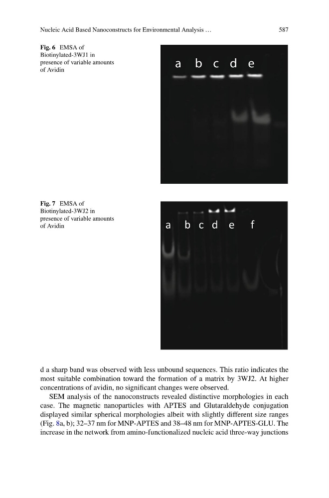
Fig. 6 EMSA of
Biotinylated-3WJ1 in
presence of variable amounts
of Avidin
a b c d e
Fig. 7 EMSA of
Biotinylated-3WJ2 in
presence of variable amounts
of Avidin
a b c d e f
d a sharp band was observed with less unbound sequences. This ratio indicates the
most suitable combination toward the formation of a matrix by 3WJ2. At higher
concentrations of avidin, no significant changes were observed.
SEM analysis of the nanoconstructs revealed distinctive morphologies in each
case. The magnetic nanoparticles with APTES and Glutaraldehyde conjugation
displayed similar spherical morphologies albeit with slightly different size ranges
(Fig. 8a, b); 32–37 nm for MNP-APTES and 38–48 nm for MNP-APTES-GLU. The
increase in the network from amino-functionalized nucleic acid three-way junctions