

550
M. Singh et al.
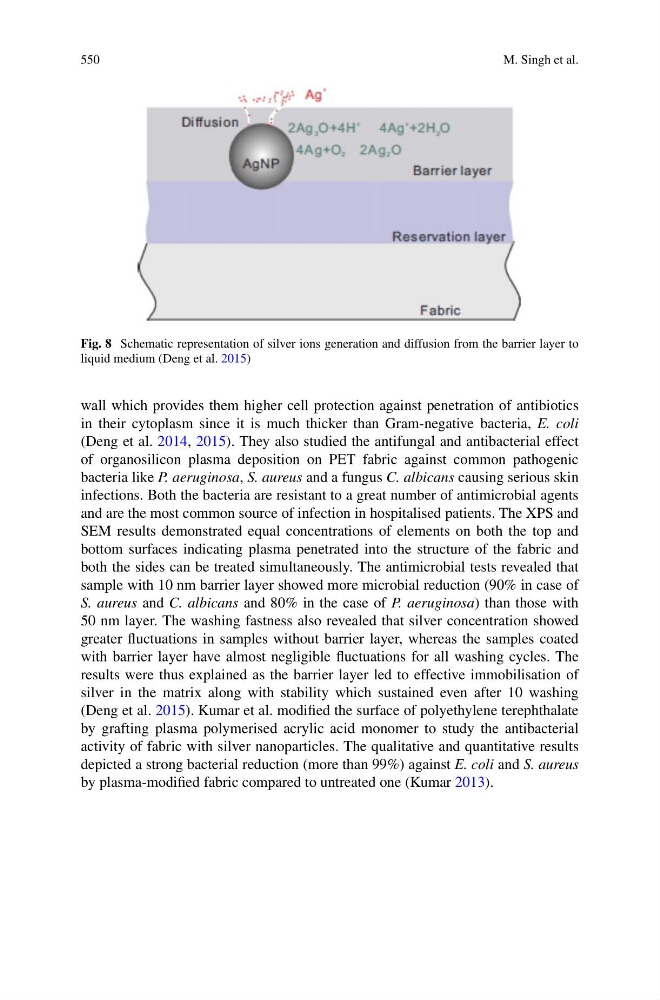
Fig. 8 Schematic representation of silver ions generation and diffusion from the barrier layer to
liquid medium (Deng et al. 2015)
wall which provides them higher cell protection against penetration of antibiotics
in their cytoplasm since it is much thicker than Gram-negative bacteria, E. coli
(Deng et al. 2014, 2015). They also studied the antifungal and antibacterial effect
of organosilicon plasma deposition on PET fabric against common pathogenic
bacteria like P. aeruginosa, S. aureus and a fungus C. albicans causing serious skin
infections. Both the bacteria are resistant to a great number of antimicrobial agents
and are the most common source of infection in hospitalised patients. The XPS and
SEM results demonstrated equal concentrations of elements on both the top and
bottom surfaces indicating plasma penetrated into the structure of the fabric and
both the sides can be treated simultaneously. The antimicrobial tests revealed that
sample with 10 nm barrier layer showed more microbial reduction (90% in case of
S. aureus and C. albicans and 80% in the case of P. aeruginosa) than those with
50 nm layer. The washing fastness also revealed that silver concentration showed
greater fluctuations in samples without barrier layer, whereas the samples coated
with barrier layer have almost negligible fluctuations for all washing cycles. The
results were thus explained as the barrier layer led to effective immobilisation of
silver in the matrix along with stability which sustained even after 10 washing
(Deng et al. 2015). Kumar et al. modified the surface of polyethylene terephthalate
by grafting plasma polymerised acrylic acid monomer to study the antibacterial
activity of fabric with silver nanoparticles. The qualitative and quantitative results
depicted a strong bacterial reduction (more than 99%) against E. coli and S. aureus
by plasma-modified fabric compared to untreated one (Kumar 2013).
M. Singh et al.
Fig. 8 Schematic representation of silver ions generation and diffusion from the barrier layer to
liquid medium (Deng et al. 2015)
wall which provides them higher cell protection against penetration of antibiotics
in their cytoplasm since it is much thicker than Gram-negative bacteria, E. coli
(Deng et al. 2014, 2015). They also studied the antifungal and antibacterial effect
of organosilicon plasma deposition on PET fabric against common pathogenic
bacteria like P. aeruginosa, S. aureus and a fungus C. albicans causing serious skin
infections. Both the bacteria are resistant to a great number of antimicrobial agents
and are the most common source of infection in hospitalised patients. The XPS and
SEM results demonstrated equal concentrations of elements on both the top and
bottom surfaces indicating plasma penetrated into the structure of the fabric and
both the sides can be treated simultaneously. The antimicrobial tests revealed that
sample with 10 nm barrier layer showed more microbial reduction (90% in case of
S. aureus and C. albicans and 80% in the case of P. aeruginosa) than those with
50 nm layer. The washing fastness also revealed that silver concentration showed
greater fluctuations in samples without barrier layer, whereas the samples coated
with barrier layer have almost negligible fluctuations for all washing cycles. The
results were thus explained as the barrier layer led to effective immobilisation of
silver in the matrix along with stability which sustained even after 10 washing
(Deng et al. 2015). Kumar et al. modified the surface of polyethylene terephthalate
by grafting plasma polymerised acrylic acid monomer to study the antibacterial
activity of fabric with silver nanoparticles. The qualitative and quantitative results
depicted a strong bacterial reduction (more than 99%) against E. coli and S. aureus
by plasma-modified fabric compared to untreated one (Kumar 2013).