

Eco-friendly Surface Modification …
549
C–C and C–H groups confirming the chain scission induced by plasma particles and
further oxidation of the carboxylate groups. Corona treatment leads to an increasing
amount of silver on PES and PA fabric although PES fabric contained higher amount
due to more number of carboxyl groups and benzene ring in their skeleton confirmed by surface-enhanced Raman spectroscopy of the strong interaction between
AgNPs and benzene ring. The bacterial reduction test showed maximum reduction
by silver-loaded polyamide fabric with 99% reduction than polyester for both the
bacteria, i.e. S. aureus and E. coli. Improved antibacterial resistance can be attributed
to enhanced surface roughness and hydrophilicity which made silver nanoparticles to
easily adhere to the fabric and in turn to bacteria to finally degrade them. The antibacterial effect was almost not altered after washing in both the silver-loaded fabrics
indicating outstanding laundering durability (Radeti´ c et al. 2008). Radetic and Ilic
also studied the antifungal effect on silver-coated, dyed and undyed corona-treated
fabrics against a most common fungus responsible for itching skin infections, i.e.
Candida albicans using disperse dyes. Antifungal tests showed more percent fungal
reduction in corona-treated PA fabric but after 5 washing the % reduction was more
in PES fabric which is in the direct effect of higher adsorption of silver nanoparticles.
The fungal reduction of dyed untreated and corona-treated PES fabric loaded with
AgNPs was more than in undyed samples which could be due to the presence of
amino groups in the dye structure which is known to obstruct the fungal growth by
cell membrane lysis (Ili´ c et al. 2009).
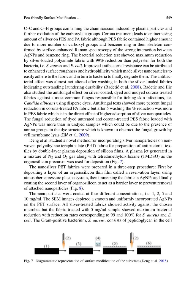
Deng et al. studied a novel method for incorporating silver nanoparticles on nonwoven polyethylene terephthalate (PET) fabric for preparation of antibacterial textiles by double-layer plasma deposition of silicon films. A plasma jet generated in
a mixture of N 2 and O 2 gas along with tetradimethyldisiloxane (TMDSO) as the
organosilicon precursor was used for deposition (Fig. 7).
The nanosilver PET fabrics were prepared in a three-step procedure: First by
depositing a layer of an organosilicon thin film called a reservation layer, using
atmospheric pressure plasma system, then immersing the fabric in AgNPs and finally
coating the second layer of organosilicon to act as a barrier layer to prevent removal
of attached nanoparticles (Fig. 8).
The nanoparticles were coated at four different concentrations, i.e. 1, 2, 5 and
10 mg/ml. The SEM images depicted a smooth and uniformly incorporated AgNPs
on the PET surface. All sliver-treated fabrics showed activity against the chosen
microbes but the fabric treated with 5 mg/ml sample showed maximum bacterial
reduction with reduction rates corresponding to 99 and 100% for S. aureus and E.
coli. The Gram-positive bacterium, S. aureus, consists of peptidoglycan in the cell
Fig. 7 Diagrammatic representation of surface modification of the substrate (Deng et al. 2015)
549
C–C and C–H groups confirming the chain scission induced by plasma particles and
further oxidation of the carboxylate groups. Corona treatment leads to an increasing
amount of silver on PES and PA fabric although PES fabric contained higher amount
due to more number of carboxyl groups and benzene ring in their skeleton confirmed by surface-enhanced Raman spectroscopy of the strong interaction between
AgNPs and benzene ring. The bacterial reduction test showed maximum reduction
by silver-loaded polyamide fabric with 99% reduction than polyester for both the
bacteria, i.e. S. aureus and E. coli. Improved antibacterial resistance can be attributed
to enhanced surface roughness and hydrophilicity which made silver nanoparticles to
easily adhere to the fabric and in turn to bacteria to finally degrade them. The antibacterial effect was almost not altered after washing in both the silver-loaded fabrics
indicating outstanding laundering durability (Radeti´ c et al. 2008). Radetic and Ilic
also studied the antifungal effect on silver-coated, dyed and undyed corona-treated
fabrics against a most common fungus responsible for itching skin infections, i.e.
Candida albicans using disperse dyes. Antifungal tests showed more percent fungal
reduction in corona-treated PA fabric but after 5 washing the % reduction was more
in PES fabric which is in the direct effect of higher adsorption of silver nanoparticles.
The fungal reduction of dyed untreated and corona-treated PES fabric loaded with
AgNPs was more than in undyed samples which could be due to the presence of
amino groups in the dye structure which is known to obstruct the fungal growth by
cell membrane lysis (Ili´ c et al. 2009).
Deng et al. studied a novel method for incorporating silver nanoparticles on nonwoven polyethylene terephthalate (PET) fabric for preparation of antibacterial textiles by double-layer plasma deposition of silicon films. A plasma jet generated in
a mixture of N 2 and O 2 gas along with tetradimethyldisiloxane (TMDSO) as the
organosilicon precursor was used for deposition (Fig. 7).
The nanosilver PET fabrics were prepared in a three-step procedure: First by
depositing a layer of an organosilicon thin film called a reservation layer, using
atmospheric pressure plasma system, then immersing the fabric in AgNPs and finally
coating the second layer of organosilicon to act as a barrier layer to prevent removal
of attached nanoparticles (Fig. 8).
The nanoparticles were coated at four different concentrations, i.e. 1, 2, 5 and
10 mg/ml. The SEM images depicted a smooth and uniformly incorporated AgNPs
on the PET surface. All sliver-treated fabrics showed activity against the chosen
microbes but the fabric treated with 5 mg/ml sample showed maximum bacterial
reduction with reduction rates corresponding to 99 and 100% for S. aureus and E.
coli. The Gram-positive bacterium, S. aureus, consists of peptidoglycan in the cell
Fig. 7 Diagrammatic representation of surface modification of the substrate (Deng et al. 2015)