

78
Algae
plates are present, the apical below the central doublet. Each plate corresponds to a flagellar constriction, and the space between them possibly contains fibrillar material; in cross-section, a stellate structure is visible, the arm of which connect with the A-Tubules of the peripheral doublets (Figure 2.41c).
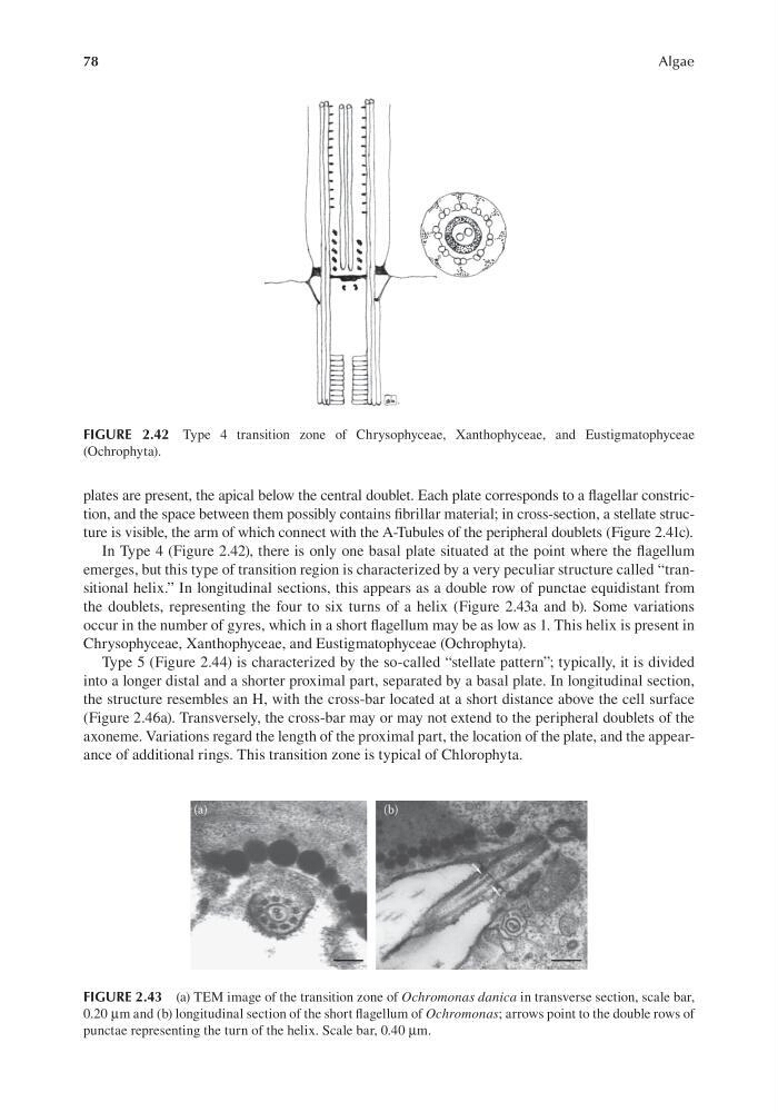
In Type 4 (Figure 2.42), there is only one basal plate situated at the point where the flagellum
emerges, but this type of transition region is characterized by a very peculiar structure called “transitional helix.” In longitudinal sections, this appears as a double row of punctae equidistant from
the doublets, representing the four to six turns of a helix (Figure 2.43a and b). Some variations
occur in the number of gyres, which in a short flagellum may be as low as 1. This helix is present in
Chrysophyceae, Xanthophyceae, and Eustigmatophyceae (Ochrophyta).
Type 5 (Figure 2.44) is characterized by the so-called “stellate pattern”; typically, it is divided
into a longer distal and a shorter proximal part, separated by a basal plate. In longitudinal section,
the structure resembles an H, with the cross-bar located at a short distance above the cell surface
(Figure 2.46a). Transversely, the cross-bar may or may not extend to the peripheral doublets of the
axoneme. Variations regard the length of the proximal part, the location of the plate, and the appearance of additional rings. This transition zone is typical of Chlorophyta.
FIGURE 2.42 Type 4 transition zone of Chrysophyceae, Xanthophyceae, and Eustigmatophyceae
(Ochrophyta).
FIGURE 2.43 (a) TEM image of the transition zone of Ochromonas danica in transverse section, scale bar,
0.20 μm and (b) longitudinal section of the short flagellum of Ochromonas; arrows point to the double rows of
punctae representing the turn of the helix. Scale bar, 0.40 μm.
Algae
plates are present, the apical below the central doublet. Each plate corresponds to a flagellar constriction, and the space between them possibly contains fibrillar material; in cross-section, a stellate structure is visible, the arm of which connect with the A-Tubules of the peripheral doublets (Figure 2.41c).
In Type 4 (Figure 2.42), there is only one basal plate situated at the point where the flagellum
emerges, but this type of transition region is characterized by a very peculiar structure called “transitional helix.” In longitudinal sections, this appears as a double row of punctae equidistant from
the doublets, representing the four to six turns of a helix (Figure 2.43a and b). Some variations
occur in the number of gyres, which in a short flagellum may be as low as 1. This helix is present in
Chrysophyceae, Xanthophyceae, and Eustigmatophyceae (Ochrophyta).
Type 5 (Figure 2.44) is characterized by the so-called “stellate pattern”; typically, it is divided
into a longer distal and a shorter proximal part, separated by a basal plate. In longitudinal section,
the structure resembles an H, with the cross-bar located at a short distance above the cell surface
(Figure 2.46a). Transversely, the cross-bar may or may not extend to the peripheral doublets of the
axoneme. Variations regard the length of the proximal part, the location of the plate, and the appearance of additional rings. This transition zone is typical of Chlorophyta.
FIGURE 2.42 Type 4 transition zone of Chrysophyceae, Xanthophyceae, and Eustigmatophyceae
(Ochrophyta).
FIGURE 2.43 (a) TEM image of the transition zone of Ochromonas danica in transverse section, scale bar,
0.20 μm and (b) longitudinal section of the short flagellum of Ochromonas; arrows point to the double rows of
punctae representing the turn of the helix. Scale bar, 0.40 μm.