

77
Anatomy
is typical of the Euglenophyceae. Among these algae, Entosiphon sulcatum is unique for its long
transition region, the spiral of which surrounds the proximal 1 μm of the central doublet.
Type 3 transition regions possess a double system of complex plates. Variations exist due to the
distance separating the plates or due to the presence of interposition material between them. In the
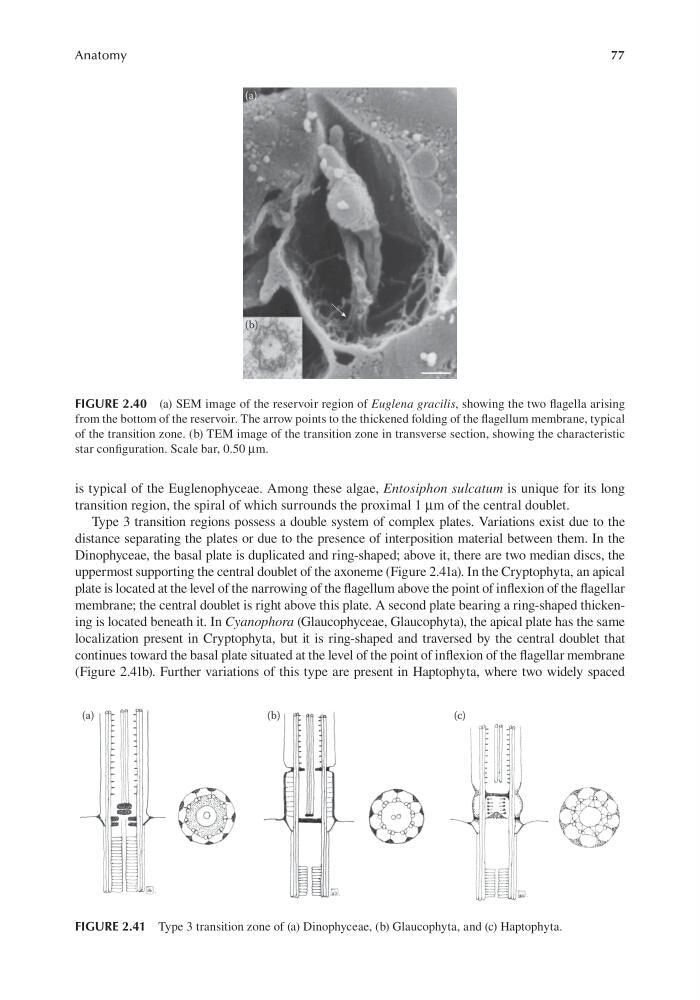
Dinophyceae, the basal plate is duplicated and ring-shaped; above it, there are two median discs, the
uppermost supporting the central doublet of the axoneme (Figure 2.41a). In the Cryptophyta, an apical
plate is located at the level of the narrowing of the flagellum above the point of inflexion of the flagellar
membrane; the central doublet is right above this plate. A second plate bearing a ring-shaped thickening is located beneath it. In Cyanophora (Glaucophyceae, Glaucophyta), the apical plate has the same
localization present in Cryptophyta, but it is ring-shaped and traversed by the central doublet that
continues toward the basal plate situated at the level of the point of inflexion of the flagellar membrane
(Figure 2.41b). Further variations of this type are present in Haptophyta, where two widely spaced
FIGURE 2.40 (a) SEM image of the reservoir region of Euglena gracilis, showing the two flagella arising
from the bottom of the reservoir. The arrow points to the thickened folding of the flagellum membrane, typical
of the transition zone. (b) TEM image of the transition zone in transverse section, showing the characteristic
star configuration. Scale bar, 0.50 μm.
FIGURE 2.41 Type 3 transition zone of (a) Dinophyceae, (b) Glaucophyta, and (c) Haptophyta.
Anatomy
is typical of the Euglenophyceae. Among these algae, Entosiphon sulcatum is unique for its long
transition region, the spiral of which surrounds the proximal 1 μm of the central doublet.
Type 3 transition regions possess a double system of complex plates. Variations exist due to the
distance separating the plates or due to the presence of interposition material between them. In the
Dinophyceae, the basal plate is duplicated and ring-shaped; above it, there are two median discs, the
uppermost supporting the central doublet of the axoneme (Figure 2.41a). In the Cryptophyta, an apical
plate is located at the level of the narrowing of the flagellum above the point of inflexion of the flagellar
membrane; the central doublet is right above this plate. A second plate bearing a ring-shaped thickening is located beneath it. In Cyanophora (Glaucophyceae, Glaucophyta), the apical plate has the same
localization present in Cryptophyta, but it is ring-shaped and traversed by the central doublet that
continues toward the basal plate situated at the level of the point of inflexion of the flagellar membrane
(Figure 2.41b). Further variations of this type are present in Haptophyta, where two widely spaced
FIGURE 2.40 (a) SEM image of the reservoir region of Euglena gracilis, showing the two flagella arising
from the bottom of the reservoir. The arrow points to the thickened folding of the flagellum membrane, typical
of the transition zone. (b) TEM image of the transition zone in transverse section, showing the characteristic
star configuration. Scale bar, 0.50 μm.
FIGURE 2.41 Type 3 transition zone of (a) Dinophyceae, (b) Glaucophyta, and (c) Haptophyta.