

74
Algae
two flagella differ in length, the rods also differ from each other in thickness and fine structure, the
longer flagellum carrying a more complex rod than the shorter flagellum. In genera with a flagellar
apparatus reduced to one long emergent flagellum and one short flagellum not extending beyond
the reservoir region, only the emergent flagellum retains the rod, which extends its entire length.
An example is Euglena gracilis; in this alga, the rod arises just above the flagellar transition zone
and is located lateroventrally with respect to the axoneme and the cell body. In thin cross-sections
of isolated and demembranated flagella, the rod appears hollow with an outer diameter of 90 nm.
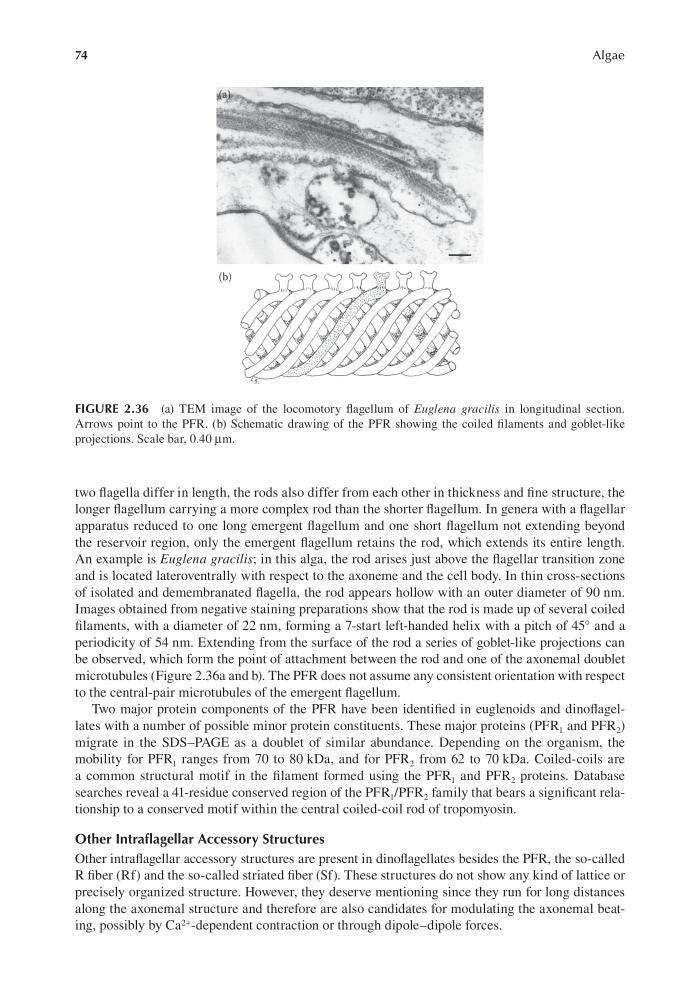
Images obtained from negative staining preparations show that the rod is made up of several coiled
filaments, with a diameter of 22 nm, forming a 7-start left-handed helix with a pitch of 45° and a
periodicity of 54 nm. Extending from the surface of the rod a series of goblet-like projections can
be observed, which form the point of attachment between the rod and one of the axonemal doublet
microtubules (Figure 2.36a and b). The PFR does not assume any consistent orientation with respect
to the central-pair microtubules of the emergent flagellum.
Two major protein components of the PFR have been identified in euglenoids and dinoflagellates with a number of possible minor protein constituents. These major proteins (PFR 1 and PFR 2 )
migrate in the SDS–PAGE as a doublet of similar abundance. Depending on the organism, the
mobility for PFR 1 ranges from 70 to 80 kDa, and for PFR 2 from 62 to 70 kDa. Coiled-coils are
a common structural motif in the filament formed using the PFR 1 and PFR 2 proteins. Database
searches reveal a 41-residue conserved region of the PFR 1 /PFR 2 family that bears a significant relationship to a conserved motif within the central coiled-coil rod of tropomyosin.
Other Intraflagellar Accessory Structures
Other intraflagellar accessory structures are present in dinoflagellates besides the PFR, the so-called
R fiber (Rf) and the so-called striated fiber (Sf). These structures do not show any kind of lattice or
precisely organized structure. However, they deserve mentioning since they run for long distances
along the axonemal structure and therefore are also candidates for modulating the axonemal beating, possibly by Ca 2+ -dependent contraction or through dipole–dipole forces.
FIGURE 2.36 (a) TEM image of the locomotory flagellum of Euglena gracilis in longitudinal section.
Arrows point to the PFR. (b) Schematic drawing of the PFR showing the coiled filaments and goblet-like
projections. Scale bar, 0.40 μm.
Algae
two flagella differ in length, the rods also differ from each other in thickness and fine structure, the
longer flagellum carrying a more complex rod than the shorter flagellum. In genera with a flagellar
apparatus reduced to one long emergent flagellum and one short flagellum not extending beyond
the reservoir region, only the emergent flagellum retains the rod, which extends its entire length.
An example is Euglena gracilis; in this alga, the rod arises just above the flagellar transition zone
and is located lateroventrally with respect to the axoneme and the cell body. In thin cross-sections
of isolated and demembranated flagella, the rod appears hollow with an outer diameter of 90 nm.
Images obtained from negative staining preparations show that the rod is made up of several coiled
filaments, with a diameter of 22 nm, forming a 7-start left-handed helix with a pitch of 45° and a
periodicity of 54 nm. Extending from the surface of the rod a series of goblet-like projections can
be observed, which form the point of attachment between the rod and one of the axonemal doublet
microtubules (Figure 2.36a and b). The PFR does not assume any consistent orientation with respect
to the central-pair microtubules of the emergent flagellum.
Two major protein components of the PFR have been identified in euglenoids and dinoflagellates with a number of possible minor protein constituents. These major proteins (PFR 1 and PFR 2 )
migrate in the SDS–PAGE as a doublet of similar abundance. Depending on the organism, the
mobility for PFR 1 ranges from 70 to 80 kDa, and for PFR 2 from 62 to 70 kDa. Coiled-coils are
a common structural motif in the filament formed using the PFR 1 and PFR 2 proteins. Database
searches reveal a 41-residue conserved region of the PFR 1 /PFR 2 family that bears a significant relationship to a conserved motif within the central coiled-coil rod of tropomyosin.
Other Intraflagellar Accessory Structures
Other intraflagellar accessory structures are present in dinoflagellates besides the PFR, the so-called
R fiber (Rf) and the so-called striated fiber (Sf). These structures do not show any kind of lattice or
precisely organized structure. However, they deserve mentioning since they run for long distances
along the axonemal structure and therefore are also candidates for modulating the axonemal beating, possibly by Ca 2+ -dependent contraction or through dipole–dipole forces.
FIGURE 2.36 (a) TEM image of the locomotory flagellum of Euglena gracilis in longitudinal section.
Arrows point to the PFR. (b) Schematic drawing of the PFR showing the coiled filaments and goblet-like
projections. Scale bar, 0.40 μm.