

69
Anatomy
(Chrysophyceae), whose flagella and cell body are closely packed with scales of very peculiar
appearance, resembling the branched structure of a tree. Examples of the second group are
Mallomonas sp. and Synura sp. (Synurophyceae); in both genera, flagellar scales are not arranged
in a regular pattern, are very small (under 300 nm), and possess different morphological types,
the most characteristic being the annular type. As the body scales, flagellar scales are produced
in deposition vesicles, extruded from the cell and brought into correct position in relation to the
other scales and the cell surface.
As said above, flagella of the Haptophyta are usually equal in length and appearance (isokont); however, members of the genus Pavlova (Pavlovophyceae) possess two markedly unequal
flagella, the anterior one is much longer than the posterior, and carrying small, dense scales in
the form of spherical or clavate knobs. These scales are often arranged in regular rows longitudinally, or can be randomly disposed on the flagellum. Scales are formed inside the Golgi
apparatus and then released into the cell surface by fusion of the plasmalemma and the cisternal
membrane.
Flagellar scales are known from many Chlorophyta, such as Prasinophytes, Chlorodendrophyceae,
Nephroselmidophyceae, and Mamiellophyceae. These algae possess nonmineralized organic scales
on their cell body and flagella, the same type of scale being rarely present on both surfaces. On the
flagella, the scales are precisely arranged in parallel longitudinal rows, sometimes in one layer, two
layers, or even three layers on top of each other. Each layer usually contains only one type of scales.
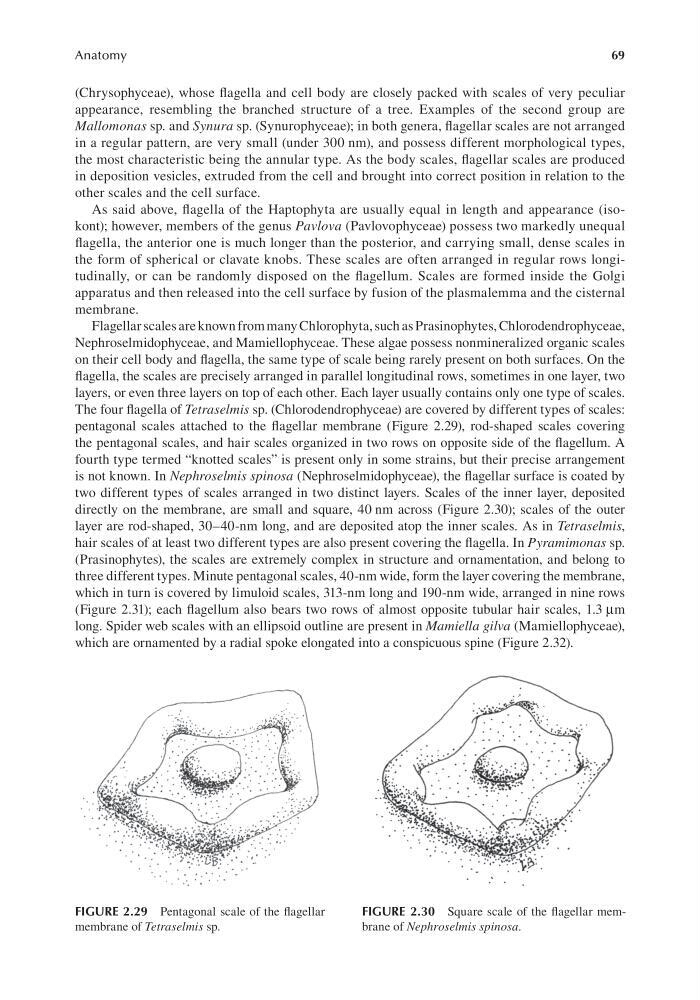
The four flagella of Tetraselmis sp. (Chlorodendrophyceae) are covered by different types of scales:
pentagonal scales attached to the flagellar membrane (Figure 2.29), rod-shaped scales covering
the pentagonal scales, and hair scales organized in two rows on opposite side of the flagellum. A
fourth type termed “knotted scales” is present only in some strains, but their precise arrangement
is not known. In Nephroselmis spinosa (Nephroselmidophyceae), the flagellar surface is coated by
two different types of scales arranged in two distinct layers. Scales of the inner layer, deposited
directly on the membrane, are small and square, 40 nm across (Figure 2.30); scales of the outer
layer are rod-shaped, 30–40-nm long, and are deposited atop the inner scales. As in Tetraselmis,
hair scales of at least two different types are also present covering the flagella. In Pyramimonas sp.
(Prasinophytes), the scales are extremely complex in structure and ornamentation, and belong to
three different types. Minute pentagonal scales, 40-nm wide, form the layer covering the membrane,
which in turn is covered by limuloid scales, 313-nm long and 190-nm wide, arranged in nine rows
(Figure 2.31); each flagellum also bears two rows of almost opposite tubular hair scales, 1.3 μm
long. Spider web scales with an ellipsoid outline are present in Mamiella gilva (Mamiellophyceae),
which are ornamented by a radial spoke elongated into a conspicuous spine (Figure 2.32).
FIGURE 2.29 Pentagonal scale of the flagellar
membrane of Tetraselmis sp.
FIGURE 2.30 Square scale of the flagellar membrane of Nephroselmis spinosa.
Anatomy
(Chrysophyceae), whose flagella and cell body are closely packed with scales of very peculiar
appearance, resembling the branched structure of a tree. Examples of the second group are
Mallomonas sp. and Synura sp. (Synurophyceae); in both genera, flagellar scales are not arranged
in a regular pattern, are very small (under 300 nm), and possess different morphological types,
the most characteristic being the annular type. As the body scales, flagellar scales are produced
in deposition vesicles, extruded from the cell and brought into correct position in relation to the
other scales and the cell surface.
As said above, flagella of the Haptophyta are usually equal in length and appearance (isokont); however, members of the genus Pavlova (Pavlovophyceae) possess two markedly unequal
flagella, the anterior one is much longer than the posterior, and carrying small, dense scales in
the form of spherical or clavate knobs. These scales are often arranged in regular rows longitudinally, or can be randomly disposed on the flagellum. Scales are formed inside the Golgi
apparatus and then released into the cell surface by fusion of the plasmalemma and the cisternal
membrane.
Flagellar scales are known from many Chlorophyta, such as Prasinophytes, Chlorodendrophyceae,
Nephroselmidophyceae, and Mamiellophyceae. These algae possess nonmineralized organic scales
on their cell body and flagella, the same type of scale being rarely present on both surfaces. On the
flagella, the scales are precisely arranged in parallel longitudinal rows, sometimes in one layer, two
layers, or even three layers on top of each other. Each layer usually contains only one type of scales.
The four flagella of Tetraselmis sp. (Chlorodendrophyceae) are covered by different types of scales:
pentagonal scales attached to the flagellar membrane (Figure 2.29), rod-shaped scales covering
the pentagonal scales, and hair scales organized in two rows on opposite side of the flagellum. A
fourth type termed “knotted scales” is present only in some strains, but their precise arrangement
is not known. In Nephroselmis spinosa (Nephroselmidophyceae), the flagellar surface is coated by
two different types of scales arranged in two distinct layers. Scales of the inner layer, deposited
directly on the membrane, are small and square, 40 nm across (Figure 2.30); scales of the outer
layer are rod-shaped, 30–40-nm long, and are deposited atop the inner scales. As in Tetraselmis,
hair scales of at least two different types are also present covering the flagella. In Pyramimonas sp.
(Prasinophytes), the scales are extremely complex in structure and ornamentation, and belong to
three different types. Minute pentagonal scales, 40-nm wide, form the layer covering the membrane,
which in turn is covered by limuloid scales, 313-nm long and 190-nm wide, arranged in nine rows
(Figure 2.31); each flagellum also bears two rows of almost opposite tubular hair scales, 1.3 μm
long. Spider web scales with an ellipsoid outline are present in Mamiella gilva (Mamiellophyceae),
which are ornamented by a radial spoke elongated into a conspicuous spine (Figure 2.32).
FIGURE 2.29 Pentagonal scale of the flagellar
membrane of Tetraselmis sp.
FIGURE 2.30 Square scale of the flagellar membrane of Nephroselmis spinosa.