

66
Algae
Second Level
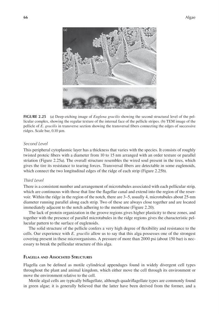
This peripheral cytoplasmic layer has a thickness that varies with the species. It consists of roughly
twisted proteic fibers with a diameter from 10 to 15 nm arranged with an order texture or parallel
striation (Figure 2.25a). The overall structure resembles the wired soul present in the tires, which
gives the tire its resistance to tearing forces. Transversal fibers are detectable in some euglenoids,
which connect the two longitudinal edges of the ridge of each strip (Figure 2.25b).
Third Level
There is a consistent number and arrangement of microtubules associated with each pellicular strip,
which are continuous with those that line the flagellar canal and extend into the region of the reservoir. Within the ridge in the region of the notch, there are 3–5, usually 4, microtubules about 25-nm
diameter running parallel along each strip. Two of these are always close together and are located
immediately adjacent to the notch adhering to the membrane (Figure 2.20).
The lack of protein organization in the groove regions gives higher plasticity to these zones, and
together with the presence of parallel microtubules in the ridge regions gives the characteristic pellucular pattern to the surface of euglenoids.
The solid structure of the pellicle confers a very high degree of flexibility and resistance to the
cells. Our experience with E. gracilis allow us to say that this alga possesses one of the strongest
covering present in these microorganisms. A pressure of more than 2000 psi (about 150 bar) is necessary to break the pellicular structure of this alga.
Flagella and associaTed sTrUcTUres
Flagella can be defined as motile cylindrical appendages found in widely divergent cell types
throughout the plant and animal kingdom, which either move the cell through its environment or
move the environment relative to the cell.
Motile algal cells are typically biflagellate, although quadriflagellate types are commonly found
in green algae; it is generally believed that the latter have been derived from the former, and a
FIGURE 2.25 (a) Deep-etching image of Euglena gracilis showing the second structural level of the pellicular complex, showing the regular texture of the internal face of the pellicle stripes. (b) TEM image of the
pellicle of E. gracilis in transverse section showing the transversal fibers connecting the edges of successive
ridges. Scale bar, 0.10 μm.
Algae
Second Level
This peripheral cytoplasmic layer has a thickness that varies with the species. It consists of roughly
twisted proteic fibers with a diameter from 10 to 15 nm arranged with an order texture or parallel
striation (Figure 2.25a). The overall structure resembles the wired soul present in the tires, which
gives the tire its resistance to tearing forces. Transversal fibers are detectable in some euglenoids,
which connect the two longitudinal edges of the ridge of each strip (Figure 2.25b).
Third Level
There is a consistent number and arrangement of microtubules associated with each pellicular strip,
which are continuous with those that line the flagellar canal and extend into the region of the reservoir. Within the ridge in the region of the notch, there are 3–5, usually 4, microtubules about 25-nm
diameter running parallel along each strip. Two of these are always close together and are located
immediately adjacent to the notch adhering to the membrane (Figure 2.20).
The lack of protein organization in the groove regions gives higher plasticity to these zones, and
together with the presence of parallel microtubules in the ridge regions gives the characteristic pellucular pattern to the surface of euglenoids.
The solid structure of the pellicle confers a very high degree of flexibility and resistance to the
cells. Our experience with E. gracilis allow us to say that this alga possesses one of the strongest
covering present in these microorganisms. A pressure of more than 2000 psi (about 150 bar) is necessary to break the pellicular structure of this alga.
Flagella and associaTed sTrUcTUres
Flagella can be defined as motile cylindrical appendages found in widely divergent cell types
throughout the plant and animal kingdom, which either move the cell through its environment or
move the environment relative to the cell.
Motile algal cells are typically biflagellate, although quadriflagellate types are commonly found
in green algae; it is generally believed that the latter have been derived from the former, and a
FIGURE 2.25 (a) Deep-etching image of Euglena gracilis showing the second structural level of the pellicular complex, showing the regular texture of the internal face of the pellicle stripes. (b) TEM image of the
pellicle of E. gracilis in transverse section showing the transversal fibers connecting the edges of successive
ridges. Scale bar, 0.10 μm.