

65
Anatomy
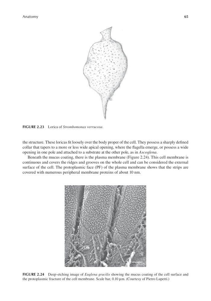
the structure. These loricas fit loosely over the body proper of the cell. They possess a sharply defined
collar that tapers to a more or less wide apical opening, where the flagella emerge, or possess a wide
opening in one pole and attached to a substrate at the other pole, as in Ascoglena.
Beneath the mucus coating, there is the plasma membrane (Figure 2.24). This cell membrane is
continuous and covers the ridges and grooves on the whole cell and can be considered the external
surface of the cell. The protoplasmic face (PF) of the plasma membrane shows that the strips are
covered with numerous peripheral membrane proteins of about 10 nm.
FIGURE 2.23 Lorica of Strombomonas verrucosa.
FIGURE 2.24 Deep-etching image of Euglena gracilis showing the mucus coating of the cell surface and
the protoplasmic fracture of the cell membrane. Scale bar, 0.10 μm. (Courtesy of Pietro Lupetti.)
Anatomy
the structure. These loricas fit loosely over the body proper of the cell. They possess a sharply defined
collar that tapers to a more or less wide apical opening, where the flagella emerge, or possess a wide
opening in one pole and attached to a substrate at the other pole, as in Ascoglena.
Beneath the mucus coating, there is the plasma membrane (Figure 2.24). This cell membrane is
continuous and covers the ridges and grooves on the whole cell and can be considered the external
surface of the cell. The protoplasmic face (PF) of the plasma membrane shows that the strips are
covered with numerous peripheral membrane proteins of about 10 nm.
FIGURE 2.23 Lorica of Strombomonas verrucosa.
FIGURE 2.24 Deep-etching image of Euglena gracilis showing the mucus coating of the cell surface and
the protoplasmic fracture of the cell membrane. Scale bar, 0.10 μm. (Courtesy of Pietro Lupetti.)