

64
Algae
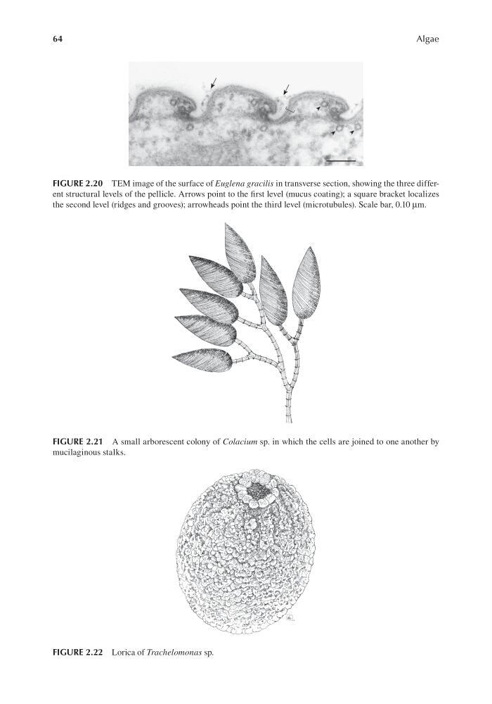
FIGURE 2.20 TEM image of the surface of Euglena gracilis in transverse section, showing the three different structural levels of the pellicle. Arrows point to the first level (mucus coating); a square bracket localizes
the second level (ridges and grooves); arrowheads point the third level (microtubules). Scale bar, 0.10 μm.
FIGURE 2.21 A small arborescent colony of Colacium sp. in which the cells are joined to one another by
mucilaginous stalks.
FIGURE 2.22 Lorica of Trachelomonas sp.
Algae
FIGURE 2.20 TEM image of the surface of Euglena gracilis in transverse section, showing the three different structural levels of the pellicle. Arrows point to the first level (mucus coating); a square bracket localizes
the second level (ridges and grooves); arrowheads point the third level (microtubules). Scale bar, 0.10 μm.
FIGURE 2.21 A small arborescent colony of Colacium sp. in which the cells are joined to one another by
mucilaginous stalks.
FIGURE 2.22 Lorica of Trachelomonas sp.