

63
Anatomy
These strips or striae can be described as long ribbons usually arising in the flagellar pocket and
extending from the cell apex to the posterior. Each strip is curved at both its edges, and in transverse
section it shows a notch, an arched, or a slightly concave ridge, a convex groove and a heel region
where adjacent strips interlock and articulate. The strips can be arranged helically or longitudinally;
the first arrangement, very elastic, is present in the “plastic euglenoids” (e.g., Euglena, Peranema,
Distigma), either heterotrophic or phototrophic, where the strips are more than 16. Their relational
sliding over one another along the articulation edges permit the cells to undergo “euglenoid movement” or “metaboly.” This movement is a sort of peristaltic movement consisting of a cytoplasmic
dilation forming at the front of the cell and passing to the rear. The return movement of the cytoplasm is brought about without dilation. The more rigid longitudinal arrangement is present in the
“aplastic euglenoids” (e.g., Petalomonas, Pleotia, Entosiphon), all heterotrophic, where the strips
are usually less than 12. These euglenids are not capable of metaboly.
The ultrastructure of the pellicular complex shows three different structural levels (Figure 2.20):
• The plasma membrane with its mucilage coating (first level)
• An electron-opaque layer organized in ridges and grooves (second level)
• The microtubular system (third level)
First Level
A dense irregular layer of mucilaginous glycoproteins covers the external surface of the cell. It has a
fuzzy texture that, however, has a somehow ordered structure of orientated threads. Mucilage bodies present beneath the cell surface secrete the mucilaginous glycoproteins. The consolidation of the
secretory products and their arrangement at one pole or round the periphery of the cell leads to the
formation of peduncles (stalks of fixation) and other enveloping structures homologs to the loricas of
Chrysophyceae and Chlorophyceae. Peduncles are present in Colacium, an euglenophyte that forms
small arborescent colonies (Figure 2.21). Its cells, with reduced flagella, are attached by their anterior
pole by a peduncle consisting of an axis of neutral polysaccharides and a cortex of acid polysaccharides. Loricas are present in Trachelomonas sp. (Figures 1.1bm and 2.22), Strombomonas verrucosa
(Figure 2.23) and Ascoglena; they are very rigid, made up of mucilaginous filaments impregnated
with ferric hydroxide or manganese compounds which confer an orange, brown to black coloration to
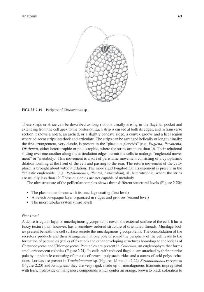
FIGURE 2.19 Periplast of Chroomonas sp.
Anatomy
These strips or striae can be described as long ribbons usually arising in the flagellar pocket and
extending from the cell apex to the posterior. Each strip is curved at both its edges, and in transverse
section it shows a notch, an arched, or a slightly concave ridge, a convex groove and a heel region
where adjacent strips interlock and articulate. The strips can be arranged helically or longitudinally;
the first arrangement, very elastic, is present in the “plastic euglenoids” (e.g., Euglena, Peranema,
Distigma), either heterotrophic or phototrophic, where the strips are more than 16. Their relational
sliding over one another along the articulation edges permit the cells to undergo “euglenoid movement” or “metaboly.” This movement is a sort of peristaltic movement consisting of a cytoplasmic
dilation forming at the front of the cell and passing to the rear. The return movement of the cytoplasm is brought about without dilation. The more rigid longitudinal arrangement is present in the
“aplastic euglenoids” (e.g., Petalomonas, Pleotia, Entosiphon), all heterotrophic, where the strips
are usually less than 12. These euglenids are not capable of metaboly.
The ultrastructure of the pellicular complex shows three different structural levels (Figure 2.20):
• The plasma membrane with its mucilage coating (first level)
• An electron-opaque layer organized in ridges and grooves (second level)
• The microtubular system (third level)
First Level
A dense irregular layer of mucilaginous glycoproteins covers the external surface of the cell. It has a
fuzzy texture that, however, has a somehow ordered structure of orientated threads. Mucilage bodies present beneath the cell surface secrete the mucilaginous glycoproteins. The consolidation of the
secretory products and their arrangement at one pole or round the periphery of the cell leads to the
formation of peduncles (stalks of fixation) and other enveloping structures homologs to the loricas of
Chrysophyceae and Chlorophyceae. Peduncles are present in Colacium, an euglenophyte that forms
small arborescent colonies (Figure 2.21). Its cells, with reduced flagella, are attached by their anterior
pole by a peduncle consisting of an axis of neutral polysaccharides and a cortex of acid polysaccharides. Loricas are present in Trachelomonas sp. (Figures 1.1bm and 2.22), Strombomonas verrucosa
(Figure 2.23) and Ascoglena; they are very rigid, made up of mucilaginous filaments impregnated
with ferric hydroxide or manganese compounds which confer an orange, brown to black coloration to
FIGURE 2.19 Periplast of Chroomonas sp.