

62
Algae
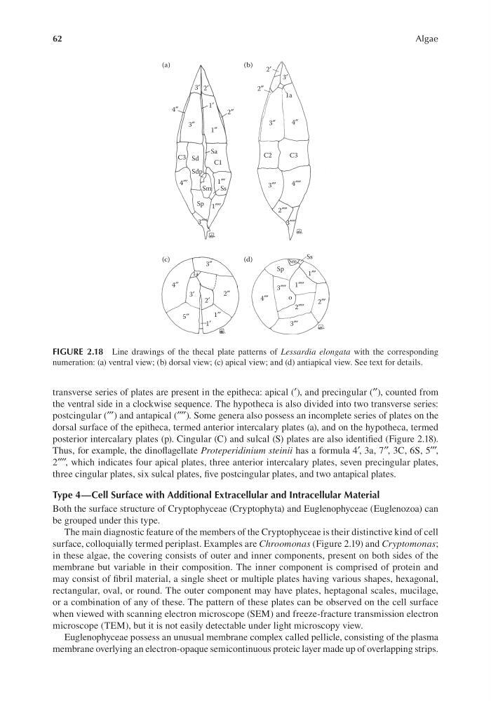
transverse series of plates are present in the epitheca: apical (′), and precingular (′′), counted from
the ventral side in a clockwise sequence. The hypotheca is also divided into two transverse series:
postcingular (′′′) and antapical (′′′′). Some genera also possess an incomplete series of plates on the
dorsal surface of the epitheca, termed anterior intercalary plates (a), and on the hypotheca, termed
posterior intercalary plates (p). Cingular (C) and sulcal (S) plates are also identified (Figure 2.18).
Thus, for example, the dinoflagellate Proteperidinium steinii has a formula 4′, 3a, 7′′, 3C, 6S, 5′′′,
2′′′′, which indicates four apical plates, three anterior intercalary plates, seven precingular plates,
three cingular plates, six sulcal plates, five postcingular plates, and two antapical plates.
Type 4—Cell Surface with Additional Extracellular and Intracellular Material
Both the surface structure of Cryptophyceae (Cryptophyta) and Euglenophyceae (Euglenozoa) can
be grouped under this type.
The main diagnostic feature of the members of the Cryptophyceae is their distinctive kind of cell
surface, colloquially termed periplast. Examples are Chroomonas (Figure 2.19) and Cryptomonas;
in these algae, the covering consists of outer and inner components, present on both sides of the
membrane but variable in their composition. The inner component is comprised of protein and
may consist of fibril material, a single sheet or multiple plates having various shapes, hexagonal,
rectangular, oval, or round. The outer component may have plates, heptagonal scales, mucilage,
or a combination of any of these. The pattern of these plates can be observed on the cell surface
when viewed with scanning electron microscope (SEM) and freeze-fracture transmission electron
microscope (TEM), but it is not easily detectable under light microscopy view.
Euglenophyceae possess an unusual membrane complex called pellicle, consisting of the plasma
membrane overlying an electron-opaque semicontinuous proteic layer made up of overlapping strips.
2′
2′
3′
(a)
(b)
(c)
(d)
3′
1′
1a
2″
2″
1″
3″
3″
4″
4″
C3
C2
C3
Sd
Sa
C1
Sdp
4″′
4″′
4″″
3″′
2″″
3″
4′′
2′ ′
5′′
1′ ′
1′
3′ 2′
1a
3″″
1″′
1″′
3″′
2″′
1″′′
1″′′
3″′′
3″′′
2″′′
o
Sm Ss
Sm
Ss
Sp
Sp
FIGURE 2.18 Line drawings of the thecal plate patterns of Lessardia elongata with the corresponding
numeration: (a) ventral view; (b) dorsal view; (c) apical view; and (d) antiapical view. See text for details.
Algae
transverse series of plates are present in the epitheca: apical (′), and precingular (′′), counted from
the ventral side in a clockwise sequence. The hypotheca is also divided into two transverse series:
postcingular (′′′) and antapical (′′′′). Some genera also possess an incomplete series of plates on the
dorsal surface of the epitheca, termed anterior intercalary plates (a), and on the hypotheca, termed
posterior intercalary plates (p). Cingular (C) and sulcal (S) plates are also identified (Figure 2.18).
Thus, for example, the dinoflagellate Proteperidinium steinii has a formula 4′, 3a, 7′′, 3C, 6S, 5′′′,
2′′′′, which indicates four apical plates, three anterior intercalary plates, seven precingular plates,
three cingular plates, six sulcal plates, five postcingular plates, and two antapical plates.
Type 4—Cell Surface with Additional Extracellular and Intracellular Material
Both the surface structure of Cryptophyceae (Cryptophyta) and Euglenophyceae (Euglenozoa) can
be grouped under this type.
The main diagnostic feature of the members of the Cryptophyceae is their distinctive kind of cell
surface, colloquially termed periplast. Examples are Chroomonas (Figure 2.19) and Cryptomonas;
in these algae, the covering consists of outer and inner components, present on both sides of the
membrane but variable in their composition. The inner component is comprised of protein and
may consist of fibril material, a single sheet or multiple plates having various shapes, hexagonal,
rectangular, oval, or round. The outer component may have plates, heptagonal scales, mucilage,
or a combination of any of these. The pattern of these plates can be observed on the cell surface
when viewed with scanning electron microscope (SEM) and freeze-fracture transmission electron
microscope (TEM), but it is not easily detectable under light microscopy view.
Euglenophyceae possess an unusual membrane complex called pellicle, consisting of the plasma
membrane overlying an electron-opaque semicontinuous proteic layer made up of overlapping strips.
2′
2′
3′
(a)
(b)
(c)
(d)
3′
1′
1a
2″
2″
1″
3″
3″
4″
4″
C3
C2
C3
Sd
Sa
C1
Sdp
4″′
4″′
4″″
3″′
2″″
3″
4′′
2′ ′
5′′
1′ ′
1′
3′ 2′
1a
3″″
1″′
1″′
3″′
2″′
1″′′
1″′′
3″′′
3″′′
2″′′
o
Sm Ss
Sm
Ss
Sp
Sp
FIGURE 2.18 Line drawings of the thecal plate patterns of Lessardia elongata with the corresponding
numeration: (a) ventral view; (b) dorsal view; (c) apical view; and (d) antiapical view. See text for details.