

60
Algae
Type 3—Cell Surface with Additional Intracellular Material in Vesicles
In this type of cell surface, the plasma is underlined by a system of flattened vesicles. An example
is the complex outer region of dinoflagellates (Dinophyceae) termed amphiesma. Beneath the cell
membrane that binds dinoflagellate motile cells, a single layer of vesicles (amphiesmal vesicles)
is almost invariably present. The vesicles may contain cellulosic plates (thecal plates) in taxa that
are thus termed thecate, or armored; or the vesicles may lack thecal plates, such taxa being termed
athecate, or unarmored or naked. In athecate taxa, the amphiesmal vesicles play a structural role. In
thecate taxa, thecal plates, one of which occurs in each vesicle, adjoin one another tightly along linear plate sutures, usually with the margin of one plate overlapping the margin of the adjacent plate.
Cellulosic plates vary from very thin to thick and can be heavily ornamented by reticula or striae;
trichocyst pores, which may lie in pits termed areolae, penetrate most of them.
A separate layer internal to the amphiesmal vesicles may develop. It is termed pellicle, though
in the case of dinoflagellates the term “pellicle” refers to a surface component completely different
from the euglenoid pellicle, hence with a completely different accepted meaning, and in our opinion
its use should be avoided. The layer consists primarily of cellulose, sometimes with a dinosporine
component, a complex organic polymer similar to sporopollenin that makes these algae fossilizable. In some athecate genera, such as Noctiluca sp., this layer forms reinforce the amphiesma, and
the cells are termed pelliculate. This layer is also sometimes present beneath the amphiesma, as in
Alexandrium sp., or Scrippsiella sp., and forms the wall of temporary cysts.
According to Dodge and Crawford (1970), the amphiesma construction falls into eight reasonably distinct categories (Figure 2.17): (1) simple membrane underlain by a single layer of vesicles
600–800 nm in length, rather flattened, circular, or irregular in shape, with a gap of at least 40 nm
between adjacent vesicles that may contain dense granular material; beneath the vesicles are parallel
rows of microtubules which lie in groups of three; this simple arrangement is present in Oxyrrhis
marina; (2) simple membrane underlain by closely packed polygonal (generally hexagonal) vesicles
0.8–1.2 μm in length, frequently containing fuzzy material; these vesicles and the cell membrane
are occasionally perforated by trichocyst pores; beneath the vesicles lie microtubules in rows of
variable number; this type of amphiesma is found in Amphidinium carterae; (3) as in category (2),
but with plug-like structures associated with the inner side of the vesicles; these plugs are cylindrical structures 120-nm long and are arranged in single lines between single or paired microtubules;
an example of this arrangement is present in Gymnodinium veneficum; (4) as in category (2), but
with thin (about 20 nm) plate-like structure in the flattened vesicles; this amphiesma characterizes Aureodinium pigmentosum; (5) in this group, the vesicles contain plates of medium thickness
(60 nm), which slightly overlap; in Woloszynskia coronata, the plates are perforated by trichocyst
pores; (6) the plates are thicker (up to 150 nm), reduced in number with a marked diversity of form;
each plate has two or more sides bearing ridges and the remaining sides have tapered flanges; where
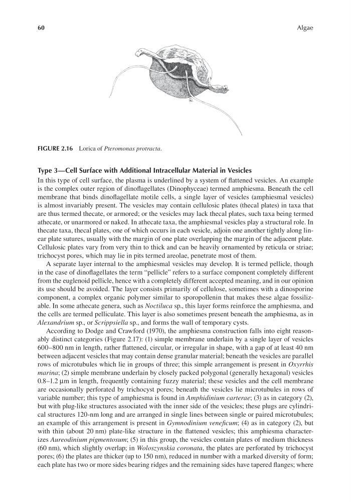
FIGURE 2.16 Lorica of Pteromonas protracta.
Algae
Type 3—Cell Surface with Additional Intracellular Material in Vesicles
In this type of cell surface, the plasma is underlined by a system of flattened vesicles. An example
is the complex outer region of dinoflagellates (Dinophyceae) termed amphiesma. Beneath the cell
membrane that binds dinoflagellate motile cells, a single layer of vesicles (amphiesmal vesicles)
is almost invariably present. The vesicles may contain cellulosic plates (thecal plates) in taxa that
are thus termed thecate, or armored; or the vesicles may lack thecal plates, such taxa being termed
athecate, or unarmored or naked. In athecate taxa, the amphiesmal vesicles play a structural role. In
thecate taxa, thecal plates, one of which occurs in each vesicle, adjoin one another tightly along linear plate sutures, usually with the margin of one plate overlapping the margin of the adjacent plate.
Cellulosic plates vary from very thin to thick and can be heavily ornamented by reticula or striae;
trichocyst pores, which may lie in pits termed areolae, penetrate most of them.
A separate layer internal to the amphiesmal vesicles may develop. It is termed pellicle, though
in the case of dinoflagellates the term “pellicle” refers to a surface component completely different
from the euglenoid pellicle, hence with a completely different accepted meaning, and in our opinion
its use should be avoided. The layer consists primarily of cellulose, sometimes with a dinosporine
component, a complex organic polymer similar to sporopollenin that makes these algae fossilizable. In some athecate genera, such as Noctiluca sp., this layer forms reinforce the amphiesma, and
the cells are termed pelliculate. This layer is also sometimes present beneath the amphiesma, as in
Alexandrium sp., or Scrippsiella sp., and forms the wall of temporary cysts.
According to Dodge and Crawford (1970), the amphiesma construction falls into eight reasonably distinct categories (Figure 2.17): (1) simple membrane underlain by a single layer of vesicles
600–800 nm in length, rather flattened, circular, or irregular in shape, with a gap of at least 40 nm
between adjacent vesicles that may contain dense granular material; beneath the vesicles are parallel
rows of microtubules which lie in groups of three; this simple arrangement is present in Oxyrrhis
marina; (2) simple membrane underlain by closely packed polygonal (generally hexagonal) vesicles
0.8–1.2 μm in length, frequently containing fuzzy material; these vesicles and the cell membrane
are occasionally perforated by trichocyst pores; beneath the vesicles lie microtubules in rows of
variable number; this type of amphiesma is found in Amphidinium carterae; (3) as in category (2),
but with plug-like structures associated with the inner side of the vesicles; these plugs are cylindrical structures 120-nm long and are arranged in single lines between single or paired microtubules;
an example of this arrangement is present in Gymnodinium veneficum; (4) as in category (2), but
with thin (about 20 nm) plate-like structure in the flattened vesicles; this amphiesma characterizes Aureodinium pigmentosum; (5) in this group, the vesicles contain plates of medium thickness
(60 nm), which slightly overlap; in Woloszynskia coronata, the plates are perforated by trichocyst
pores; (6) the plates are thicker (up to 150 nm), reduced in number with a marked diversity of form;
each plate has two or more sides bearing ridges and the remaining sides have tapered flanges; where
FIGURE 2.16 Lorica of Pteromonas protracta.