

59
Anatomy
is mainly cellulosic, while in the Chlorophyceae (Chlorophyta) xylose, mannose, and chitin may
also be present in addition to cellulose. Some members of the Chlorophyceae (Chlorophyta) and
Charophyceae (Charophyta) have calcified walls.
Lorica
These enveloping structures are present in some members of the class Chrysophyceae (Ochrophyta)
such as Dinobryon sp., or Chrysococcus sp., and in some genera of the Chlorophyceae, such as
Phacotus, Pteromonas, and Dysmorphococcus. These loricas are vase-shaped structure with a
more or less wide apical opening, where the flagella emerge. These structures can be colorless, or
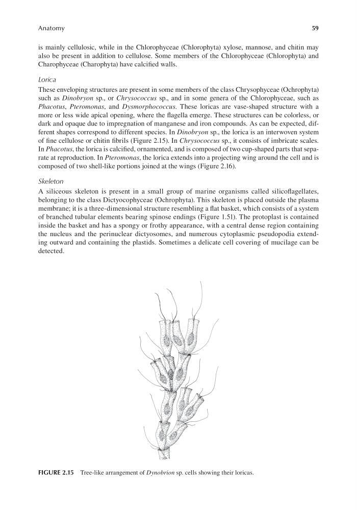
dark and opaque due to impregnation of manganese and iron compounds. As can be expected, different shapes correspond to different species. In Dinobryon sp., the lorica is an interwoven system
of fine cellulose or chitin fibrils (Figure 2.15). In Chrysococcus sp., it consists of imbricate scales.
In Phacotus, the lorica is calcified, ornamented, and is composed of two cup-shaped parts that separate at reproduction. In Pteromonas, the lorica extends into a projecting wing around the cell and is
composed of two shell-like portions joined at the wings (Figure 2.16).
Skeleton
A siliceous skeleton is present in a small group of marine organisms called silicoflagellates,
belonging to the class Dictyocophyceae (Ochrophyta). This skeleton is placed outside the plasma
membrane; it is a three-dimensional structure resembling a flat basket, which consists of a system
of branched tubular elements bearing spinose endings (Figure 1.51). The protoplast is contained
inside the basket and has a spongy or frothy appearance, with a central dense region containing
the nucleus and the perinuclear dictyosomes, and numerous cytoplasmic pseudopodia extending outward and containing the plastids. Sometimes a delicate cell covering of mucilage can be
detected.
FIGURE 2.15 Tree-like arrangement of Dynobrion sp. cells showing their loricas.
Anatomy
is mainly cellulosic, while in the Chlorophyceae (Chlorophyta) xylose, mannose, and chitin may
also be present in addition to cellulose. Some members of the Chlorophyceae (Chlorophyta) and
Charophyceae (Charophyta) have calcified walls.
Lorica
These enveloping structures are present in some members of the class Chrysophyceae (Ochrophyta)
such as Dinobryon sp., or Chrysococcus sp., and in some genera of the Chlorophyceae, such as
Phacotus, Pteromonas, and Dysmorphococcus. These loricas are vase-shaped structure with a
more or less wide apical opening, where the flagella emerge. These structures can be colorless, or
dark and opaque due to impregnation of manganese and iron compounds. As can be expected, different shapes correspond to different species. In Dinobryon sp., the lorica is an interwoven system
of fine cellulose or chitin fibrils (Figure 2.15). In Chrysococcus sp., it consists of imbricate scales.
In Phacotus, the lorica is calcified, ornamented, and is composed of two cup-shaped parts that separate at reproduction. In Pteromonas, the lorica extends into a projecting wing around the cell and is
composed of two shell-like portions joined at the wings (Figure 2.16).
Skeleton
A siliceous skeleton is present in a small group of marine organisms called silicoflagellates,
belonging to the class Dictyocophyceae (Ochrophyta). This skeleton is placed outside the plasma
membrane; it is a three-dimensional structure resembling a flat basket, which consists of a system
of branched tubular elements bearing spinose endings (Figure 1.51). The protoplast is contained
inside the basket and has a spongy or frothy appearance, with a central dense region containing
the nucleus and the perinuclear dictyosomes, and numerous cytoplasmic pseudopodia extending outward and containing the plastids. Sometimes a delicate cell covering of mucilage can be
detected.
FIGURE 2.15 Tree-like arrangement of Dynobrion sp. cells showing their loricas.