

91
Anatomy
Cercozoa—Chlorarachniophyceae
The single flagellum of the ovoid zoospores of Chlorarachion reptans possesses a root system that
consists of a microtubular component appearing as a 3 + 1 structure near the level of emergence
of the flagellum, soon increasing to 8 + 1, and a second root with a homogeneous substructure that
occupies a distinct concavity in the nuclear envelope.
In contrast to C. reptans, cells of Bigelowiella natans are basically biflagellate. The second
flagellum, however, is exceptionally short and is represented only by a barren basal body inserted
at approximately right angle to the emergent flagellum. In this alga, the four flagellar roots are
represented by microtubular structures only with no cross-banded roots. A microtubular root is
present on either sides of the long flagellum basal body; the largest and most conspicuous root,
which attaches to the outside of the long flagellum basal body, is five-stranded and forms an “L” in
the area between the nucleus and the plasma membrane. Just before terminating, this root becomes
two-stranded. The second microtubular root associated with the long flagellum basal body emerges
from the corner between the two basal bodies and is one-stranded. The third flagellar root emerges
on the outside of the short basal body and it is three-stranded. The four microtubular roots are the
most unusual; they are one-stranded and seem to emerge within the lumen to the short basal body
next to the cartwheel structure (Figure 2.57).
Mizozoa—Dinophyceae
The root apparatus of these algae is quite complex for number and appearance of ancillary
structures associated to the microtubular roots and for the spatial relationship between roots
and other cell organelles. Minor features can be considered species-specific, whereas major
components are common to almost all the dinoflagellates. The longitudinal basal body and the
transverse basal body are interconnected by a small striated connective band. A multimembered
microtubular root, the longitudinal root, originates on the left side of the longitudinal basal body
and continues posteriorly along the sulcus. A cross-striated fibrous root, the transverse striated
root, emanates from the left side of the transverse basal body and runs along the transverse flagellar canal and the cingulum. Striated connectives link the transverse striated root to both the
longitudinal root, and the longitudinal basal body, and the proximal portion of the longitudinal
AF
MA
DB
PR
BPR
SSB
BSB
MP
PF
BPR
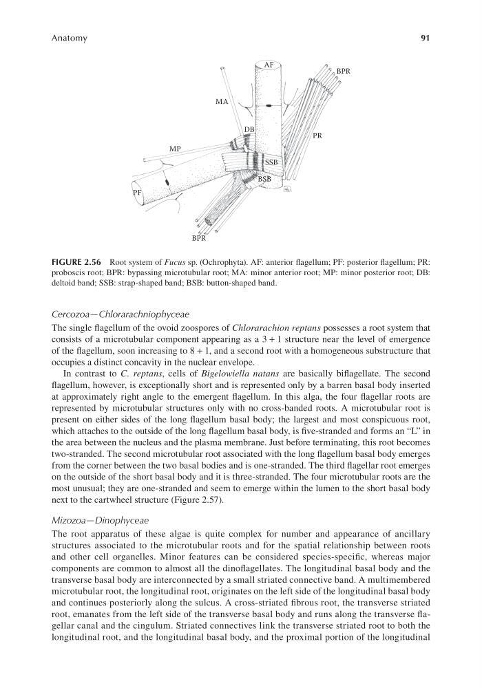
FIGURE 2.56 Root system of Fucus sp. (Ochrophyta). AF: anterior flagellum; PF: posterior flagellum; PR:
proboscis root; BPR: bypassing microtubular root; MA: minor anterior root; MP: minor posterior root; DB:
deltoid band; SSB: strap-shaped band; BSB: button-shaped band.
Anatomy
Cercozoa—Chlorarachniophyceae
The single flagellum of the ovoid zoospores of Chlorarachion reptans possesses a root system that
consists of a microtubular component appearing as a 3 + 1 structure near the level of emergence
of the flagellum, soon increasing to 8 + 1, and a second root with a homogeneous substructure that
occupies a distinct concavity in the nuclear envelope.
In contrast to C. reptans, cells of Bigelowiella natans are basically biflagellate. The second
flagellum, however, is exceptionally short and is represented only by a barren basal body inserted
at approximately right angle to the emergent flagellum. In this alga, the four flagellar roots are
represented by microtubular structures only with no cross-banded roots. A microtubular root is
present on either sides of the long flagellum basal body; the largest and most conspicuous root,
which attaches to the outside of the long flagellum basal body, is five-stranded and forms an “L” in
the area between the nucleus and the plasma membrane. Just before terminating, this root becomes
two-stranded. The second microtubular root associated with the long flagellum basal body emerges
from the corner between the two basal bodies and is one-stranded. The third flagellar root emerges
on the outside of the short basal body and it is three-stranded. The four microtubular roots are the
most unusual; they are one-stranded and seem to emerge within the lumen to the short basal body
next to the cartwheel structure (Figure 2.57).
Mizozoa—Dinophyceae
The root apparatus of these algae is quite complex for number and appearance of ancillary
structures associated to the microtubular roots and for the spatial relationship between roots
and other cell organelles. Minor features can be considered species-specific, whereas major
components are common to almost all the dinoflagellates. The longitudinal basal body and the
transverse basal body are interconnected by a small striated connective band. A multimembered
microtubular root, the longitudinal root, originates on the left side of the longitudinal basal body
and continues posteriorly along the sulcus. A cross-striated fibrous root, the transverse striated
root, emanates from the left side of the transverse basal body and runs along the transverse flagellar canal and the cingulum. Striated connectives link the transverse striated root to both the
longitudinal root, and the longitudinal basal body, and the proximal portion of the longitudinal
AF
MA
DB
PR
BPR
SSB
BSB
MP
PF
BPR
FIGURE 2.56 Root system of Fucus sp. (Ochrophyta). AF: anterior flagellum; PF: posterior flagellum; PR:
proboscis root; BPR: bypassing microtubular root; MA: minor anterior root; MP: minor posterior root; DB:
deltoid band; SSB: strap-shaped band; BSB: button-shaped band.