

92
Algae
basal body to the longitudinal root. The most distinct connective is a large electron-opaque fiber
termed the nuclear fibrous connective, which links the proximal parts of the longitudinal root,
the transverse striated root, and the transverse and longitudinal basal bodies with the nucleus.
Fibrous rings with cross-striations, called fibrous collars, encircle the flagellar canals, the longitudinal striated collar less conspicuous than the transverse striated collar. A nonstriated fiber
interconnects the two collars. The fibrous elements of this complex root apparatus are likely to
contain centrin (Figure 2.58).
Euglenozoa—Euglenophyceae
Euglenoids show a rather uniform root structure, with one microtubular root opposite each basal
body and a single microtubular root in between, termed dorsal, intermediate, and ventral roots
(Figure 2.59). The dorsal root is anchored to the dorsal basal body at the side furthest from the
ventral basal body; both the intermediate and ventral roots are associated with the ventral basal
body at its dorsal and ventral side, respectively. In Euglena mutabilis, the dorsal and the intermediate roots consist of three microtubules, while the ventral root consists of five microtubules; in
E. gracilis, the ventral root is formed by five microtubules as in E. mutabilis, while the microtubules of the dorsal root are more numerous. These roots extend from the basal bodies along the
reservoir and into the cytoplasm, usually along the cell periphery, but in some species toward
the nucleus. Flagellar roots are believed to play an important role in maintaining cell shape. In
some species, the two flagellar basal bodies are connected by a conspicuous transversely striated
connective.
Besides the intraflagellar accessory structures described above, euglenoids with two emergent
flagella possess extra-flagellar accessory structures, the so-called rootlets. Two major classes of
rootlets have been described: microtubular rootlets and filamentous rootlets. Microtubular rootlets are made of a usually complex and species-specific network (geometry, number of elements)
anchoring the flagellar apparatus in the whole cell body. However, due to known functions of microtubules in other systems, these microtubules could not only play a role of an anchor, but also provide
a stable oriented network along which cargos could be transported from inside the cell body to the
flagellar apparatus.
The filamentous rootlets are made of filaments that can contain centrin. These appendages are
attached to the pair of basal bodies located at the base of the flagella and extend inside the cytoplasm, usually in the direction of the nucleus or along the plasma membrane. Classically, they have
been considered as anchoring structures providing the role of roots to the whole flagellar apparatus.
However, they are also contractile structures, which may show transversal striations (centrin type,
not assembling type) whose periodicity varies with their contraction state.
F1
R4
R1
BBB
R2
R3
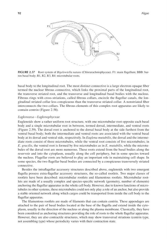
FIGURE 2.57 Root system of Bigelowiella natans (Chlorarachniophyceae). F1: main flagellum; BBB: barren basal body; R1, R2, R3, R4: microtubular roots.
Algae
basal body to the longitudinal root. The most distinct connective is a large electron-opaque fiber
termed the nuclear fibrous connective, which links the proximal parts of the longitudinal root,
the transverse striated root, and the transverse and longitudinal basal bodies with the nucleus.
Fibrous rings with cross-striations, called fibrous collars, encircle the flagellar canals, the longitudinal striated collar less conspicuous than the transverse striated collar. A nonstriated fiber
interconnects the two collars. The fibrous elements of this complex root apparatus are likely to
contain centrin (Figure 2.58).
Euglenozoa—Euglenophyceae
Euglenoids show a rather uniform root structure, with one microtubular root opposite each basal
body and a single microtubular root in between, termed dorsal, intermediate, and ventral roots
(Figure 2.59). The dorsal root is anchored to the dorsal basal body at the side furthest from the
ventral basal body; both the intermediate and ventral roots are associated with the ventral basal
body at its dorsal and ventral side, respectively. In Euglena mutabilis, the dorsal and the intermediate roots consist of three microtubules, while the ventral root consists of five microtubules; in
E. gracilis, the ventral root is formed by five microtubules as in E. mutabilis, while the microtubules of the dorsal root are more numerous. These roots extend from the basal bodies along the
reservoir and into the cytoplasm, usually along the cell periphery, but in some species toward
the nucleus. Flagellar roots are believed to play an important role in maintaining cell shape. In
some species, the two flagellar basal bodies are connected by a conspicuous transversely striated
connective.
Besides the intraflagellar accessory structures described above, euglenoids with two emergent
flagella possess extra-flagellar accessory structures, the so-called rootlets. Two major classes of
rootlets have been described: microtubular rootlets and filamentous rootlets. Microtubular rootlets are made of a usually complex and species-specific network (geometry, number of elements)
anchoring the flagellar apparatus in the whole cell body. However, due to known functions of microtubules in other systems, these microtubules could not only play a role of an anchor, but also provide
a stable oriented network along which cargos could be transported from inside the cell body to the
flagellar apparatus.
The filamentous rootlets are made of filaments that can contain centrin. These appendages are
attached to the pair of basal bodies located at the base of the flagella and extend inside the cytoplasm, usually in the direction of the nucleus or along the plasma membrane. Classically, they have
been considered as anchoring structures providing the role of roots to the whole flagellar apparatus.
However, they are also contractile structures, which may show transversal striations (centrin type,
not assembling type) whose periodicity varies with their contraction state.
F1
R4
R1
BBB
R2
R3
FIGURE 2.57 Root system of Bigelowiella natans (Chlorarachniophyceae). F1: main flagellum; BBB: barren basal body; R1, R2, R3, R4: microtubular roots.